Copyright
©2013 Baishideng Publishing Group Co.
World J Gastroenterol. Aug 14, 2013; 19(30): 4907-4916
Published online Aug 14, 2013. doi: 10.3748/wjg.v19.i30.4907
Published online Aug 14, 2013. doi: 10.3748/wjg.v19.i30.4907
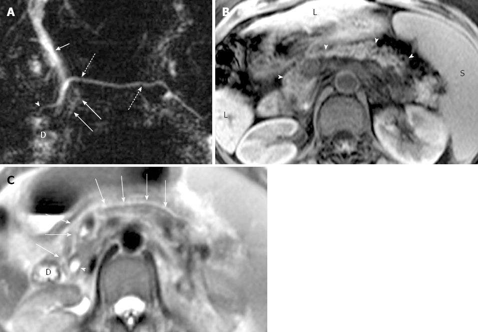
Figure 3 Magnetic resonance cholangiopancreatography and magnetic resonance imaging of incidental chronic pancreatitis in a 47-year-old woman with a history of liver cirrhosis.
A: Coronal oblique thick-section rapid acquisition with relaxation enhancement magnetic resonance (RARE-MR) cholangiogram [infinite/1100 (effective), 40-mm section thickness] of the pancreaticobiliary ducts shows slight dilatation with irregularity and strictures in the main pancreatic duct (dotted arrows) and in the duct of Santorini (arrowhead) just before entering the duodenum (D) via the minor papilla. Several side branch ectasia (long arrows) arising from the ventral duct and a normal common bile duct (short arrows) are demonstrated; B: Axial T1WI with fat saturation shows severe atrophy of the entire pancreas parenchyma (arrowheads). The liver (L) cirrhosis and splenomegaly (S) are noted; C: Axial T2WI shows main pancreatic duct (solid arrows) continued by duct of Santorini entering the duodenum (D) anterior to the common bile duct (arrowhead).
- Citation: Wang DB, Yu J, Fulcher AS, Turner MA. Pancreatitis in patients with pancreas divisum: Imaging features at MRI and MRCP. World J Gastroenterol 2013; 19(30): 4907-4916
- URL: https://www.wjgnet.com/1007-9327/full/v19/i30/4907.htm
- DOI: https://dx.doi.org/10.3748/wjg.v19.i30.4907


