Copyright
©2012 Baishideng Publishing Group Co.
World J Gastroenterol. Aug 21, 2012; 18(31): 4102-4117
Published online Aug 21, 2012. doi: 10.3748/wjg.v18.i31.4102
Published online Aug 21, 2012. doi: 10.3748/wjg.v18.i31.4102
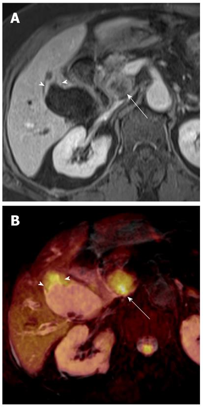
Figure 9 Gallbladder carcinoma (focal wall-thickening type) in a 77-year-old woman.
A: Axial contrast-enhanced, fat-saturated, T1-weighted image shows focal wall thickening (arrowheads) in the gallbladder with a metastatic lymph node in the portocaval space (arrow); B: Fusion image of T2-weighted image and DWI at b = 800 s/mm2 shows focal, asymmetric high signal intensity in the fundal portion of the gallbladder (arrowheads) with a hyperintense metastatic lymph node in the portocaval space (arrow). DWI: Diffusion-weighted magnetic resonance imaging.
- Citation: Lee NK, Kim S, Kim GH, Kim DU, Seo HI, Kim TU, Kang DH, Jang HJ. Diffusion-weighted imaging of biliopancreatic disorders: Correlation with conventional magnetic resonance imaging. World J Gastroenterol 2012; 18(31): 4102-4117
- URL: https://www.wjgnet.com/1007-9327/full/v18/i31/4102.htm
- DOI: https://dx.doi.org/10.3748/wjg.v18.i31.4102


