Copyright
©2011 Baishideng Publishing Group Co.
World J Gastroenterol. Sep 28, 2011; 17(36): 4090-4098
Published online Sep 28, 2011. doi: 10.3748/wjg.v17.i36.4090
Published online Sep 28, 2011. doi: 10.3748/wjg.v17.i36.4090
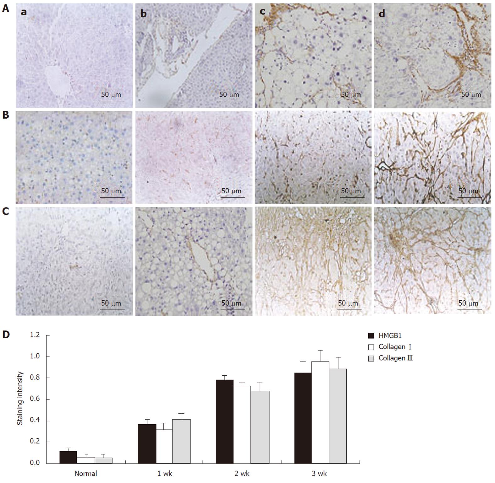
Figure 1 High-mobility group box 1 protein was upregulated after dimethylnitrosamine injection.
A: Immunohistochemical study of high-mobility group box 1 (HMGB1) distribution and expression in liver fibrosis specimens (original magnification, × 400). Brown color displays the positive expression. a: There was no immunoreactivity in the normal liver tissue; b: Weak staining in liver fibrosis tissue at 1 wk after the first dimethylnitrosamine (DMN) injection; c: Moderate staining in liver fibrosis tissue at 2 wk after the first DMN injection; d: Strong staining in liver fibrosis tissue at 3 wk after the first DMN injection; B: Immunohistochemical study of collagen type Iin liver fibrosis specimens (original magnification, × 400). Brown color displays the positive expression. Collagen type I was markedly increased during liver fibrogenesis; C: Immunohistochemical study of collagen type III in liver fibrosis specimens (original magnification, × 400). Brown color displays the positive expression. Collagen type III was markedly increased during liver fibrogenesis; D: The amount of HMGB1, collagen types I and III staining in liver tissue was measured using an image analyzer during liver fibrosis. HMGB1 was markedly increased during liver fibrogenesis, correlated with the expression of collagen types I and III (r = 0.90, P < 0.05 and r = 0.89, P < 0.05).
- Citation: Ge WS, Wu JX, Fan JG, Wang YJ, Chen YW. Inhibition of high-mobility group box 1 expression by siRNA in rat hepatic stellate cells. World J Gastroenterol 2011; 17(36): 4090-4098
- URL: https://www.wjgnet.com/1007-9327/full/v17/i36/4090.htm
- DOI: https://dx.doi.org/10.3748/wjg.v17.i36.4090


