Copyright
©2011 Baishideng Publishing Group Co.
World J Gastroenterol. Apr 21, 2011; 17(15): 1947-1960
Published online Apr 21, 2011. doi: 10.3748/wjg.v17.i15.1947
Published online Apr 21, 2011. doi: 10.3748/wjg.v17.i15.1947
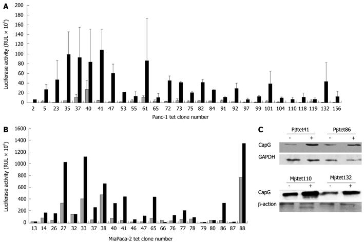
Figure 2 Identification of stable pN1βactin-rtTA2S-M2-IRES-EGFP- expressing Panc-1 (A) and MiaPaca (B) cell clones and their functionality (C).
Panc-1 and MiaPaca cells were transfected with pN1βactin-rtTA2S-M2-IRES-EGFP and stable expressors selected using 300 μg/mL G418. Clones were isolated and transfected with pTRE2hygluc, used as an indirect measure of rtTA activity. Cells were treated for 24 h with 500 ng/mL doxycycline (black bars) or an equivalent volume of PBS (white bars) and luciferase activity measured in 50 μg protein (1 mg/mL protein; 50 μL was assayed per sample). A: 90 G418 resistant clones were isolated from Panc-1 cells, of which 25% showed > 5-fold increase in luciferase activity in the stimulated (black bars in Figure 1) compared to the unstimulated (white bars in Figure 1) condition; B: Of 170 G418-resistant cells identified in MiaPaCa-2 cells, 60% showed > 5-fold increase in luciferase expression; C: Two stable pN1βactin-rtTA2S-M2-IRES-EGFP Panc-1 (Pβtet41 and Pβtet86) and MiaPaCa-2 (Mβtet110and Mβtet132) cell lines were transiently transfected with the pTRE2hygCapG vector and treated for 24 h with or without 500 ng/mL doxycycline. Cell lysates was subjected to Western Blotting for the detection of CapG. β-actin and GAPDH were used as loading controls, respectively. Tet: Tetracycline.
- Citation: Tonack S, Patel S, Jalali M, Nedjadi T, Jenkins RE, Goldring C, Neoptolemos J, Costello E. Tetracycline-inducible protein expression in pancreatic cancer cells: Effects of CapG overexpression. World J Gastroenterol 2011; 17(15): 1947-1960
- URL: https://www.wjgnet.com/1007-9327/full/v17/i15/1947.htm
- DOI: https://dx.doi.org/10.3748/wjg.v17.i15.1947


