Copyright
©2010 Baishideng Publishing Group Co.
World J Gastroenterol. Nov 7, 2010; 16(41): 5203-5210
Published online Nov 7, 2010. doi: 10.3748/wjg.v16.i41.5203
Published online Nov 7, 2010. doi: 10.3748/wjg.v16.i41.5203
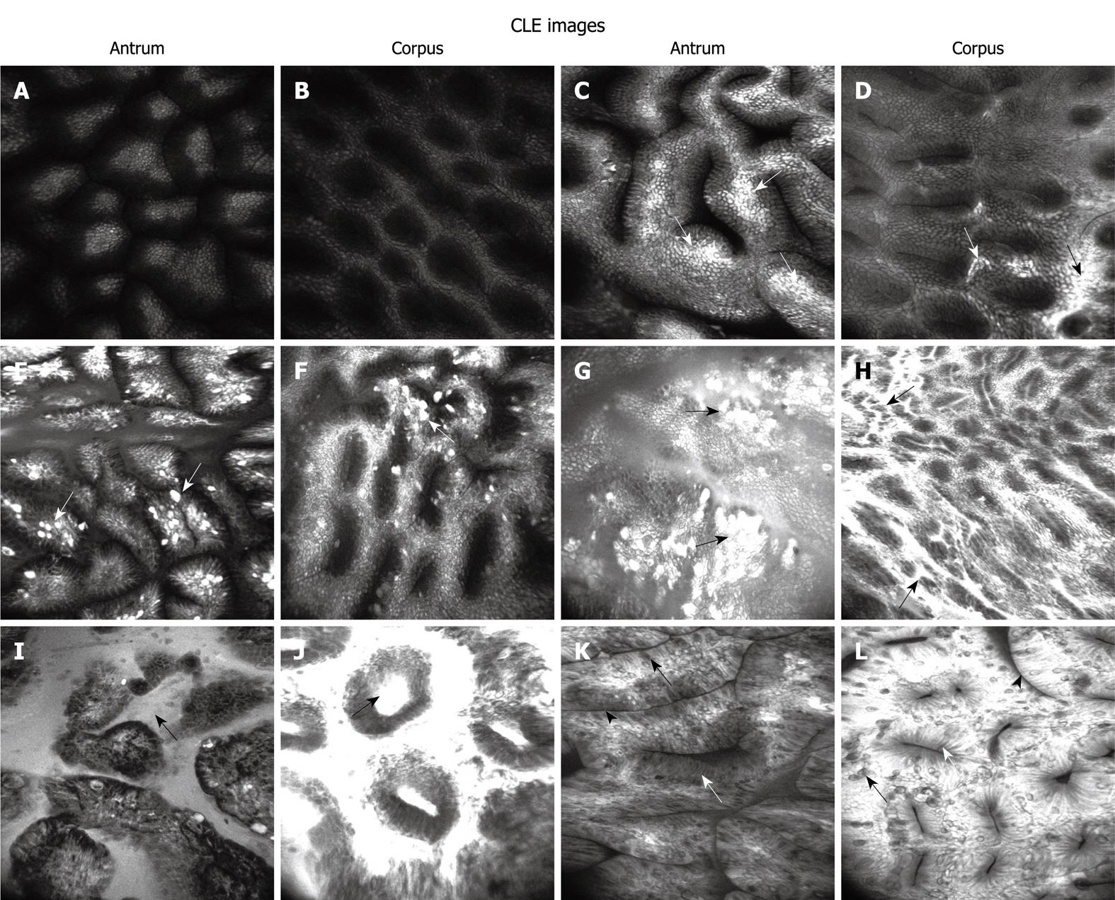
Figure 1 Confocal laser endomicroscopy classification of Helicobacter pylori-associated gastritis severity in gastric antrum and corpus.
A, B: Normal mucosa with normal antral and corporal pits, and free of fluorescein leakage; C, D: Active inflammation (mild) with slightly distorted pits and intact epithelium, scattered focal fluorescein leakage (arrows); E, F: Active inflammation (moderate) with more distorted pits and partly destroyed epithelium (arrows), and more fluorescein leakage; G, H: Active inflammation (marked) with markedly distorted pits and dilated opening, destroyed epithelium (arrows), and widespread fluorescein leakage; I, J: Glandular atrophy with decreased gastric pits and markedly dilated opening (arrows); K, L: Intestinal metaplasia with villous-like gastric pits and goblet cells (black arrows), absorptive cells (white arrows) and brush border (arrowheads) appearing. CLE: Confocal laser endomicroscopy.
-
Citation: Wang P, Ji R, Yu T, Zuo XL, Zhou CJ, Li CQ, Li Z, Li YQ. Classification of histological severity of
Helicobacter pylori -associated gastritis by confocal laser endomicroscopy. World J Gastroenterol 2010; 16(41): 5203-5210 - URL: https://www.wjgnet.com/1007-9327/full/v16/i41/5203.htm
- DOI: https://dx.doi.org/10.3748/wjg.v16.i41.5203


