Copyright
©2009 The WJG Press and Baishideng.
World J Gastroenterol. Mar 21, 2009; 15(11): 1319-1330
Published online Mar 21, 2009. doi: 10.3748/wjg.15.1319
Published online Mar 21, 2009. doi: 10.3748/wjg.15.1319
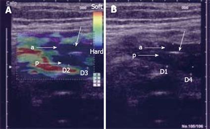
Figure 4 Elastography image acquired transabdominally in a patient with CD and stenosis of the terminal ileum.
In the left image, the bowel wall is thickened without stratification and the anterior (A) and posterior (B) bowel wall is separated by luminal air (unmarked arrow). In the right elastography image, the stenotic area is colored blue relative to the surrounding tissue, which indicates that it is hard. The patient was operated upon and the histology confirmed a fibrotic stenosis. D1: 9.5 mm; D2: 8.8 mm; D3: 10.0 mm; D4: 13.0 mm.
- Citation: Nylund K, Ødegaard S, Hausken T, Folvik G, Lied GA, Viola I, Hauser H, Gilja OH. Sonography of the small intestine. World J Gastroenterol 2009; 15(11): 1319-1330
- URL: https://www.wjgnet.com/1007-9327/full/v15/i11/1319.htm
- DOI: https://dx.doi.org/10.3748/wjg.15.1319


