Copyright
©2008 The WJG Press and Baishideng.
World J Gastroenterol. May 7, 2008; 14(17): 2740-2747
Published online May 7, 2008. doi: 10.3748/wjg.14.2740
Published online May 7, 2008. doi: 10.3748/wjg.14.2740
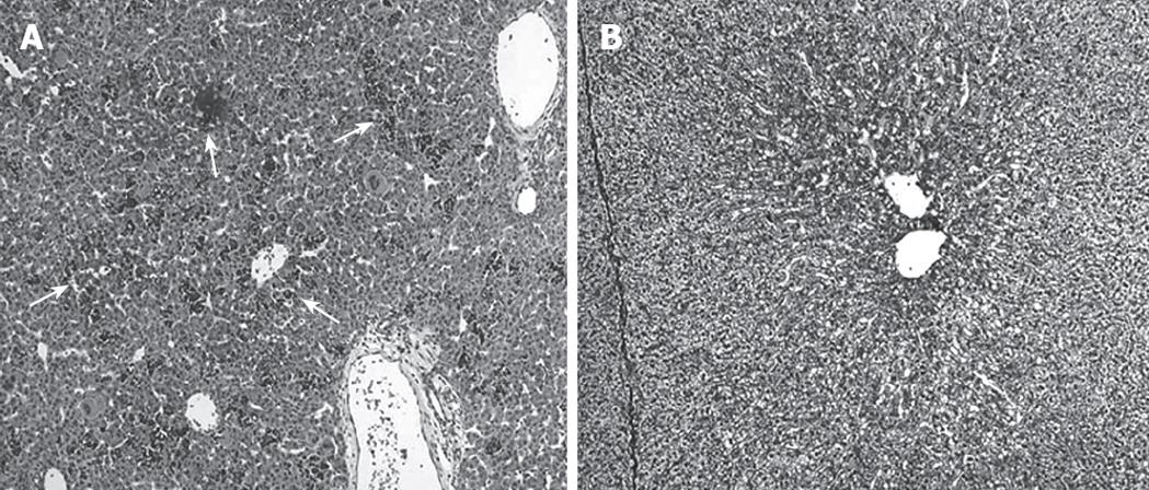
Figure 2 Histological findings of fetal porcine liver (HE staining).
A: Extramedullary hematopoiesis was observed (arrow) and the cells are largely immature at embryonic d 35 (× 100); B: A definite lobular structures and a decrease of extramedullary hematopoiesis were observed at embryonic d 56 (× 100).
- Citation: Ishii Y, Saito R, Marushima H, Ito R, Sakamoto T, Yanaga K. Hepatic reconstruction from fetal porcine liver cells using a radial flow bioreactor. World J Gastroenterol 2008; 14(17): 2740-2747
- URL: https://www.wjgnet.com/1007-9327/full/v14/i17/2740.htm
- DOI: https://dx.doi.org/10.3748/wjg.14.2740


