Copyright
©2006 Baishideng Publishing Group Co.
World J Gastroenterol. Sep 21, 2006; 12(35): 5735-5738
Published online Sep 21, 2006. doi: 10.3748/wjg.v12.i35.5735
Published online Sep 21, 2006. doi: 10.3748/wjg.v12.i35.5735
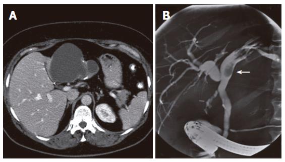
Figure 1 Abdominal CT-scan showing a large cystic mass in the left liver lobe with internal septations and calcifications in the cyst wall (A) and ERCP showing a polypoid lesion in the left hepatic duct (arrow) in case 1 (B).
- Citation: Erdogan D, Busch OR, Rauws EA, Delden OMV, Gouma DJ, Gulik TMV. Obstructive jaundice due to hepatobiliary cystadenoma or cystadenocarcinoma. World J Gastroenterol 2006; 12(35): 5735-5738
- URL: https://www.wjgnet.com/1007-9327/full/v12/i35/5735.htm
- DOI: https://dx.doi.org/10.3748/wjg.v12.i35.5735


