Copyright
©2006 Baishideng Publishing Group Co.
World J Gastroenterol. Apr 28, 2006; 12(16): 2510-2516
Published online Apr 28, 2006. doi: 10.3748/wjg.v12.i16.2510
Published online Apr 28, 2006. doi: 10.3748/wjg.v12.i16.2510
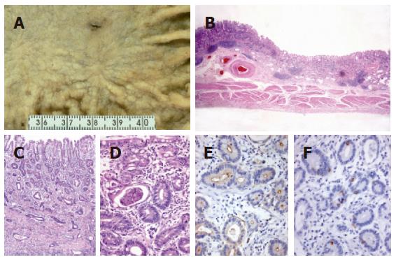
Figure 2 An early lesion of extremely well-differentiated adenocarcinoma of the stomach, complete intestinal-type (case 8).
A: Macroscopic view showing a shallow depressed lesion with an irregular margin; B: carcinoma invasion to the submucosal layer (low-power view); C and D: carcinoma mimicking the intestinal metaplasia of complete-type with basally located small nuclei, eosinophilic cytoplasm and scattered goblet cells. Note the irregular arrangement of glands and intraluminal debris; E: CD10 positivity of carcinomatous glands along the luminal surfaces; and F: MUC2 positivity of scattered goblet cells.
- Citation: Yao T, Utsunomiya T, Oya M, Nishiyama K, Tsuneyoshi M. Extremely well-differentiated adenocarcinoma of the stomach: Clinicopathological and immunohistochemical features. World J Gastroenterol 2006; 12(16): 2510-2516
- URL: https://www.wjgnet.com/1007-9327/full/v12/i16/2510.htm
- DOI: https://dx.doi.org/10.3748/wjg.v12.i16.2510


