Copyright
©2006 Baishideng Publishing Group Co.
World J Gastroenterol. Apr 21, 2006; 12(15): 2388-2393
Published online Apr 21, 2006. doi: 10.3748/wjg.v12.i15.2388
Published online Apr 21, 2006. doi: 10.3748/wjg.v12.i15.2388
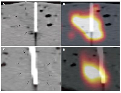
Figure 5 It shows an example of two isodense lesions on CT alone and combined PET/CT with the inserted biopsy needles.
The lesions margins are barely seen on CT images alone (fine black arrows, A and C). In comparison, the true extent and localisation of the lesion are well shown on the combined PET/CT images. Based on poor visibility on CT alone, the lesion in the bottom row was nearly missed (D).
- Citation: Veit P, Kuehle C, Beyer T, Kuehl H, Bockisch A, Antoch G. Accuracy of combined PET/CT in image-guided interventions of liver lesions: An ex-vivo study. World J Gastroenterol 2006; 12(15): 2388-2393
- URL: https://www.wjgnet.com/1007-9327/full/v12/i15/2388.htm
- DOI: https://dx.doi.org/10.3748/wjg.v12.i15.2388


