Copyright
©2005 Baishideng Publishing Group Inc.
World J Gastroenterol. Nov 21, 2005; 11(43): 6828-6832
Published online Nov 21, 2005. doi: 10.3748/wjg.v11.i43.6828
Published online Nov 21, 2005. doi: 10.3748/wjg.v11.i43.6828
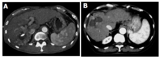
Figure 1 CT images of scattered recurrences after RFA.
A: Recurrences around the ablated tumor after RFA (case 10 in Table 2); B: Multiple recurrences scattered over the whole liver after RFA (case 6 in Table 2). The white arrows indicate the ablated area without enhancement by contrast medium, and the black arrowheads indicate scattered recurrence with enhancement.
- Citation: Kotoh K, Enjoji M, Arimura E, Morizono S, Kohjima M, Sakai H, Nakamuta M. Scattered and rapid intrahepatic recurrences after radio frequency ablation for hepatocellular carcinoma. World J Gastroenterol 2005; 11(43): 6828-6832
- URL: https://www.wjgnet.com/1007-9327/full/v11/i43/6828.htm
- DOI: https://dx.doi.org/10.3748/wjg.v11.i43.6828


