Copyright
©The Author(s) 2003.
World J Gastroenterol. Sep 15, 2003; 9(9): 2045-2049
Published online Sep 15, 2003. doi: 10.3748/wjg.v9.i9.2045
Published online Sep 15, 2003. doi: 10.3748/wjg.v9.i9.2045
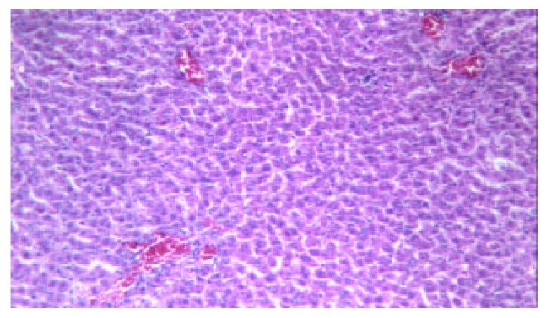
Figure 1 Light microscopy for control liver tissue, normal liver histology.
H&E × 100.
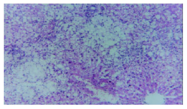
Figure 2 Light microscopy for liver tissue from a 12-week treated rat in NASH group, severe macrovesicular steatosis with mixed parenchymal inflammation and spotty focal necrosis.
H&E × 100.
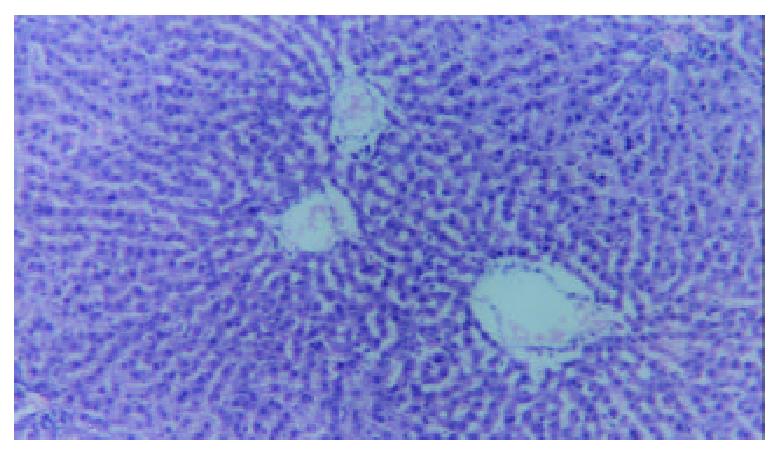
Figure 3 Light microscopy for liver tissue from a rat treated with LCD during 12-week experiment, the pathological changes of liver were obviously improved compared with the NASH group.
H&E × 100.
- Citation: Fan JG, Zhong L, Xu ZJ, Tia LY, Ding XD, Li MS, Wang GL. Effects of low-calorie diet on steatohepatitis in rats with obesity and hyperlipidemia. World J Gastroenterol 2003; 9(9): 2045-2049
- URL: https://www.wjgnet.com/1007-9327/full/v9/i9/2045.htm
- DOI: https://dx.doi.org/10.3748/wjg.v9.i9.2045


