Copyright
©The Author(s) 2003.
World J Gastroenterol. Jun 15, 2003; 9(6): 1227-1230
Published online Jun 15, 2003. doi: 10.3748/wjg.v9.i6.1227
Published online Jun 15, 2003. doi: 10.3748/wjg.v9.i6.1227
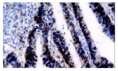
Figure 1 VEGF expression in colorectal cancer specimen.
VEGF immunoreactivity was observed mainly in the cytoplasm of tumor cells, and also frequently in stromal cells.
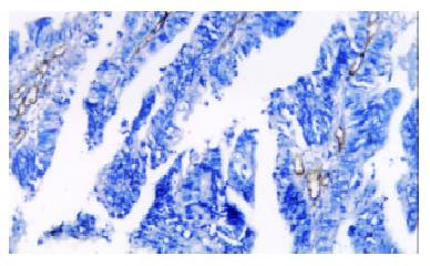
Figure 2 Microvascular density in colorectal cancer specimen.
The single brown-stained cell indicates an endothelial cell that was stained for the presence of CD34.
- Citation: Zheng S, Han MY, Xiao ZX, Peng JP, Dong Q. Clinical significance of vascular endothelial growth factor expression and neovascularization in colorectal carcinoma. World J Gastroenterol 2003; 9(6): 1227-1230
- URL: https://www.wjgnet.com/1007-9327/full/v9/i6/1227.htm
- DOI: https://dx.doi.org/10.3748/wjg.v9.i6.1227


