Copyright
©The Author(s) 2003.
World J Gastroenterol. May 15, 2003; 9(5): 1114-1118
Published online May 15, 2003. doi: 10.3748/wjg.v9.i5.1114
Published online May 15, 2003. doi: 10.3748/wjg.v9.i5.1114
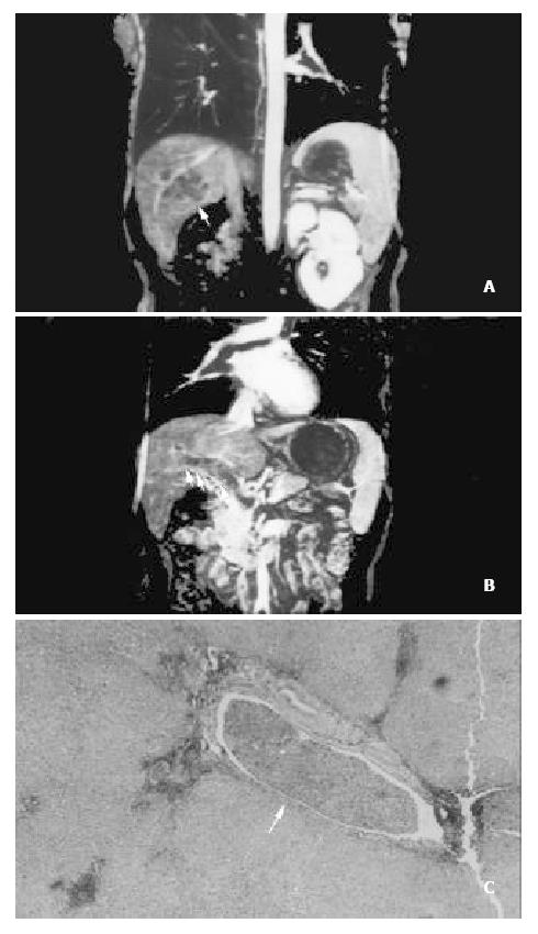
Figure 1 A patient with hepatocellular carcinoma in the right lobe.
(A) Source image of 3-dimensional contrast-enhanced MR angiography demonstrates a hypointense tumor in the right lobe (arrow). (B) Multiplanar reconstruction of 3-dimensional contrast-enhanced MR angiography shows widened main and right portal vein with filling defects (arrow). (C) The patient underwent tumor resection and removal of tumor thrombi in the portal vein. Histopathology (HE × 40) reveals tumor throm-bus in the portal vein (arrow).
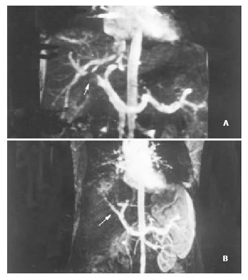
Figure 2 A patient with hepatocellular carcinoma in the right robe.
(A) 3-dimensional contrast-enhanced MR angiography depicts a nodular filling defect in the right portal vein (arrow).
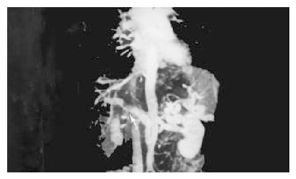
Figure 3 A patient with hepatocellular carcinoma in the left lobe.
3-dimensional contrast-enhanced MR angiography demonstrates occluded left portal vein and irregular filling defect in the main portal vein (arrow). The right portal vein is normal.
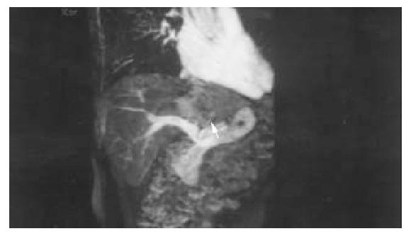
Figure 4 A patient with hepatocellualr carcinoma in the left lobe (arrow).
The left portal vein is not visualized and interpreted as occlusion. At surgery, however, it was only com-pressed by the tumor.
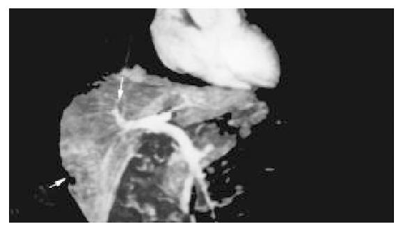
Figure 5 A patient with hepatocellular carcinoma in the right lobe (thick arrow).
The normal right anterior portal vein (thin arrow) is misinterpreted as the right portal vein. But at surgery, the right posterior portal vein was involved by the tumor.
- Citation: Lin J, Zhou KR, Chen ZW, Wang JH, Wu ZQ, Fan J. Three-dimensional contrast-enhanced MR angiography in diagnosis of portal vein involvement by hepatic tumors. World J Gastroenterol 2003; 9(5): 1114-1118
- URL: https://www.wjgnet.com/1007-9327/full/v9/i5/1114.htm
- DOI: https://dx.doi.org/10.3748/wjg.v9.i5.1114


