Copyright
©The Author(s) 2003.
World J Gastroenterol. Nov 15, 2003; 9(11): 2605-2608
Published online Nov 15, 2003. doi: 10.3748/wjg.v9.i11.2605
Published online Nov 15, 2003. doi: 10.3748/wjg.v9.i11.2605
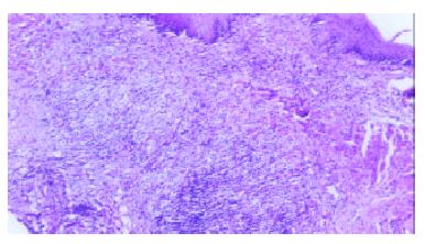
Figure 1 In the experimental group, on the 5th day after the procedure, muscle layers of the rat esophagus exhibited an inflammatory reaction.
H&E stain, × 4.
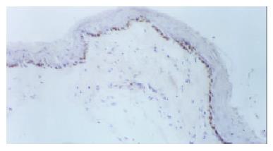
Figure 2 In the experimental group, on the 5th day after the procedure, the basal cells of the squamous epithelium in rat esophagus exhibited strong PCNA expression.
Immunostaining, × 4.
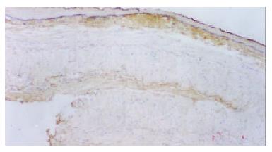
Figure 3 In the experimental group, on the 30th day after the procedure, the substratum of the rat esophageal mucosa and muscle layers exhibited strong FN expression.
H&E stain, × 4.
- Citation: Cheng YS, Li MH, Yang RJ, Zhang HZ, Ding ZX, Zhuang QX, Jiang ZM, Shang KZ. Restenosis following balloon dilation of benign esophageal stenosis. World J Gastroenterol 2003; 9(11): 2605-2608
- URL: https://www.wjgnet.com/1007-9327/full/v9/i11/2605.htm
- DOI: https://dx.doi.org/10.3748/wjg.v9.i11.2605


