Copyright
©The Author(s) 2003.
World J Gastroenterol. Nov 15, 2003; 9(11): 2509-2512
Published online Nov 15, 2003. doi: 10.3748/wjg.v9.i11.2509
Published online Nov 15, 2003. doi: 10.3748/wjg.v9.i11.2509
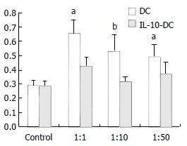
Figure 1 Inhibitory effect of IL-10-DC on allogenic T cell proliferation.
aP < 0.05, bP < 0.01, vs DC group.
Figure 2 Flow cytometric analysis for apoptotic T cells induced by IL-10-DC.
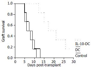
Figure 3 Survival (%) of recipient small intestines from control group (n = 6), untransduced DC group (n = 6) and IL-10-DC group (n = 6).
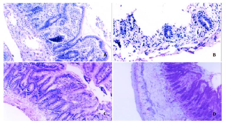
Figure 4 Histological comparison of allografts between control group rats and IL-10-DC pretreated rats.
Control group showed acute rejection sign on POD 3 (A) and mucosal sloughing and necrosis and destruction of normal glandular architecture on POD 7 (B). IL-10-DC group demonstrated mild lymphocyte infiltration and blunting of villi with edema on POD 7 (C) and fibroblastic proliferation extending from the submucosa to the lamina muscularis (D). (hematoxylin-erosin staining, × 200).
- Citation: Zhu M, Wei MF, Liu F, Shi HF, Wang G. Interleukin-10 modified dendritic cells induce allo-hyporesponsiveness and prolong small intestine allograft survival. World J Gastroenterol 2003; 9(11): 2509-2512
- URL: https://www.wjgnet.com/1007-9327/full/v9/i11/2509.htm
- DOI: https://dx.doi.org/10.3748/wjg.v9.i11.2509


