Copyright
©The Author(s) 2003.
World J Gastroenterol. Nov 15, 2003; 9(11): 2484-2489
Published online Nov 15, 2003. doi: 10.3748/wjg.v9.i11.2484
Published online Nov 15, 2003. doi: 10.3748/wjg.v9.i11.2484
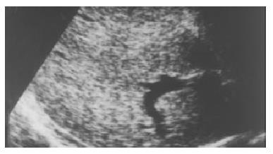
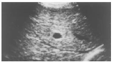
Figure 1 Active cirrhosis.
Ultrasonography showed that the hepatic parenchymal echo became coarse and dot-shaped, and the distribution was still homogenous.
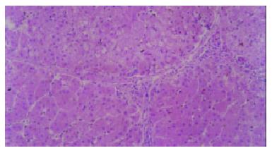
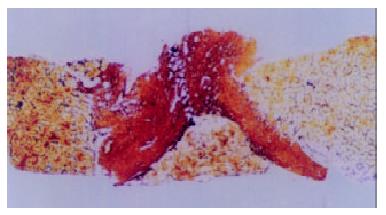
Figure 2 Active cirrhosis.
Small and sparse fibroitc septa were shown on histology. (HE stain × 100).
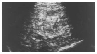
Figure 3 Chronic viral hepatitis.
On ultrasonography, the hepatic parenchymal echo became coarse and heterogeneous with speckled hyper- and hypoechoic areas.
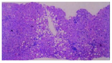
Figure 4 Chronic viral hepatitis.
On histology, focal fatty degeneration foci were shown. (HE stain, × 40).
Figure 5 Chronic viral hepatitis.
Ultrasonography showed the hepatic parenchymal echo to be strip-shaped and coarse.
Figure 6 Chronic viral hepatitis.
Wide and compact fibrotic septum was shown histologically. (Reticular fiber stain, × 40).
- Citation: Zheng RQ, Wang QH, Lu MD, Xie SB, Ren J, Su ZZ, Cai YK, Yao JL. Liver fibrosis in chronic viral hepatitis: An ultrasonographic study. World J Gastroenterol 2003; 9(11): 2484-2489
- URL: https://www.wjgnet.com/1007-9327/full/v9/i11/2484.htm
- DOI: https://dx.doi.org/10.3748/wjg.v9.i11.2484


