Copyright
©The Author(s) 2002.
World J Gastroenterol. Oct 15, 2002; 8(5): 863-867
Published online Oct 15, 2002. doi: 10.3748/wjg.v8.i5.863
Published online Oct 15, 2002. doi: 10.3748/wjg.v8.i5.863
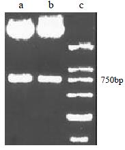
Figure 1 Restriction map of HCV E2-scFv by Sfi I/Not I digestion.
A, B: HCV E2-scFv; C: DNA Marker.
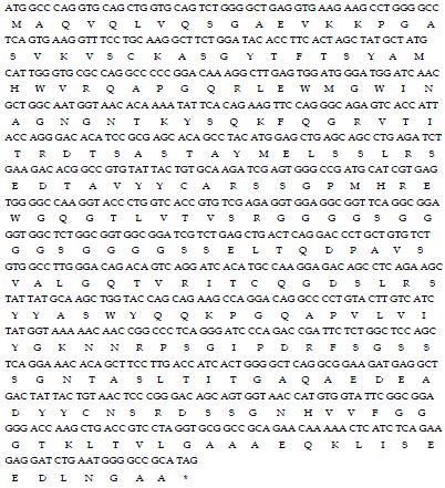
Figure 2 Nucleic acid and deduced amino acid sequences of scFv for HCV E2 protein GenBank accession number for this sequence is AF317001.
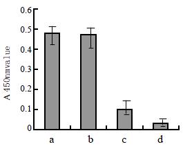
Figure 3 Absorbances of HCV-E2-scFv binding to E2 antigen by ELISA.
(a). supernant from induced XL1-blue transformed with pCANTAB5E-E2-scFv; (b). posive control; (c). supernant from non-induced XL1-blue transformed with pCANTAB5E-E2-scFv, (d). negtive control.
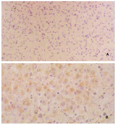
Figure 4 A.
Immunohistochemistry of liver tissue from health person; B. Immunohistochemistry of liver tissue from patients with chronic hepatitis C E2 antigen was detected in the cytoplasm of some liver cells.
- Citation: Zhong YW, Cheng J, Wang G, Shi SS, Li L, Zhang LX, Chen JM. Preparation of human single chain Fv antibody against hepatitis C virus E2 protein and its identification in immunohistochemistry. World J Gastroenterol 2002; 8(5): 863-867
- URL: https://www.wjgnet.com/1007-9327/full/v8/i5/863.htm
- DOI: https://dx.doi.org/10.3748/wjg.v8.i5.863


