Copyright
©The Author(s) 2001.
World J Gastroenterol. Apr 15, 2001; 7(2): 238-242
Published online Apr 15, 2001. doi: 10.3748/wjg.v7.i2.238
Published online Apr 15, 2001. doi: 10.3748/wjg.v7.i2.238
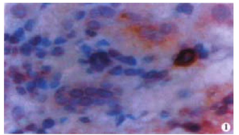
Figure 1 Oval cell identified by c-kit staining (Immunohistochemistry ABC method; original magnification: × 400)
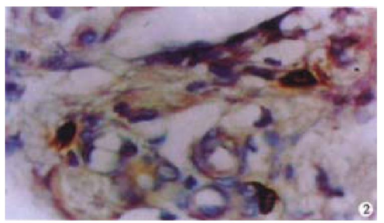
Figure 2 Oval cells were located predominantly in the periportal region in hepatic cihrrosis (Immunohistochemistry ABC method; stained by c-kit; original magnification: × 400)
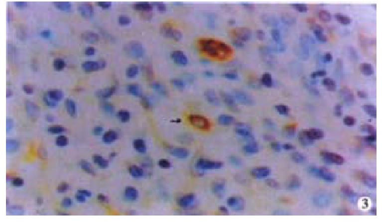
Figure 3 Oval cells were often found in close association with inflammatory cells in chronic active hepatitis (Figure 3).
(Immunohistochemistry ABC method; stained by π -GST, original magnification: × 400)
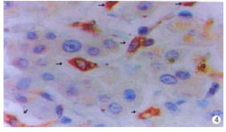
Figure 4 “Transitional cells” from a patient with hepatic cirrhosis (Immunohistochemistry ABC method; stained by CK19; original magnification: × 400)
- Citation: Ma X, Qiu DK, Peng YS. Immunohistochemical study of hepatic oval cells in human chronic viral hepatitis. World J Gastroenterol 2001; 7(2): 238-242
- URL: https://www.wjgnet.com/1007-9327/full/v7/i2/238.htm
- DOI: https://dx.doi.org/10.3748/wjg.v7.i2.238


