
















Copyright: ©Author(s) 2026.
World J Gastroenterol. Jan 21, 2027; 33(3): 123673
Published online Jan 21, 2027. doi: 10.3748/wjg.123673
Published online Jan 21, 2027. doi: 10.3748/wjg.123673
Figure 1 High METTL1 expression is associated with poor clinical prognosis of pancreatic cancer.
A: Immunohistochemical staining and quan
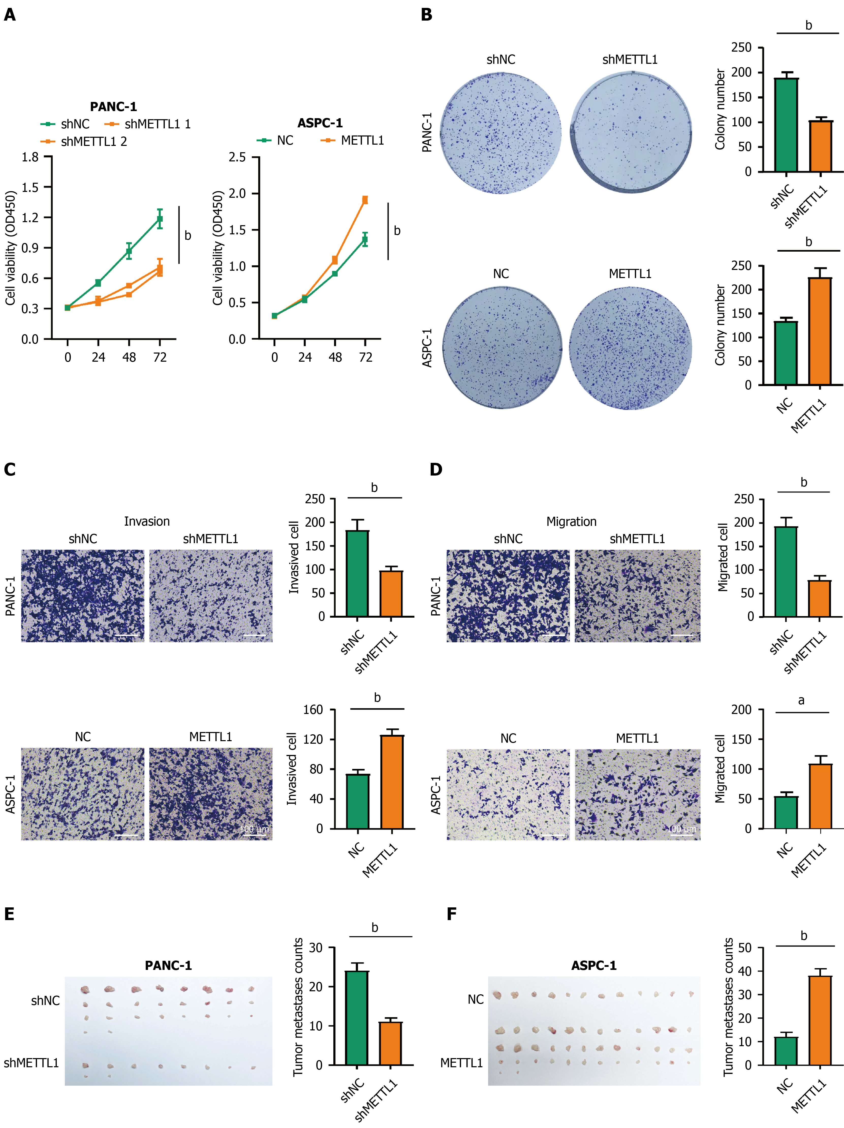
Figure 2 METTL1 promotes proliferation, invasion and in vivo peritoneal metastasis of pancreatic adenocarcinoma cells.
A: Cell Counting Kit-8 assay detecting cell proliferative activity; B: Colony formation ability and statistical colony number; C and D: Transwell migration and invasion assays and quantitative analysis of cellular motility and invasiveness; E and F: Morphological observation and nodule statistics of nude mouse intraperitoneal metastasis model for in vivo colonization evaluation. Scale bar: 100 μm; aP < 0.05, bP < 0.01.
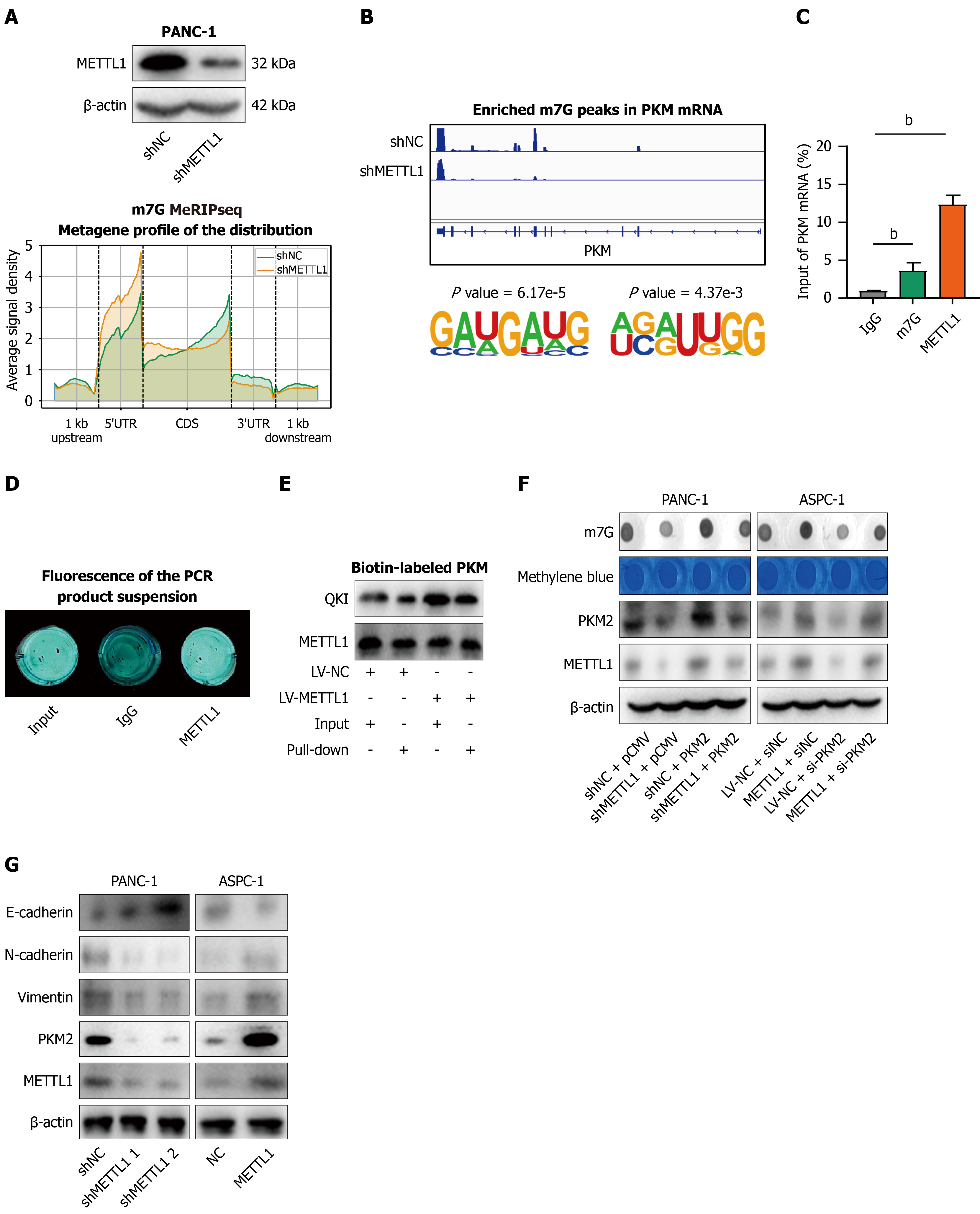
Figure 3 METTL1 regulates PKM2 expression by targeting N7-methylguanosine modification of PKM mRNA.
A: Western blot verification of METTL1 knockdown efficiency in PANC-1 cells; B: Distribution characteristics and sequence conservation of N7-methylguanosine (m7G) modification peaks on PKM mRNA before and after METTL1 knockdown via m7G-methylated RNA immunoprecipitation (MeRIP)-sequencing; C: MeRIP-quantitative polymerase chain reaction quantification of m7G modification enrichment of PKM mRNA upon METTL1 overexpression; D and E: Fluorescence imaging and biotin-labeled probe pull-down assay verifying direct binding between METTL1 and PKM mRNA; F: Changes of m7G level and PKM2 expression after METTL1 intervention detected by Dot blot and western blot; G: Western blot analysis of epithelial-mesenchymal transition biomarker expression regulated by METTL1-PKM2 axis. bP < 0.01. m7G: N7-methylguanosine; MeRIP: Methylated RNA immunoprecipitation; PCR: Polymerase chain reaction; IgG: Immunoglobulin G.
Figure 4 METTL1-PKM2 axis modulates glycolysis and lactate production and mediates H4K5 lactylation.
A: Overall glycolytic level detected by extracellular acidification rate assay; B: Lactate secretion level in cell supernatant measured by detection kit; C: Expression profiles of multiple histone lactylation modifications and specific alteration of H4K5 lactylation (H4K5 La); D and E: H4K5 La enrichment in Snail-1 promoter region analyzed by Cleavage Under Targets & Tagmentation sequencing (PRJNA1392586) and visualization; F-H: Expression changes of H4K5 La and Snail-1 after METTL1 and PKM2 intervention validated by quantitative polymerase chain reaction and western blot; I: Immunohistochemistry and immunofluorescence analysis reveals high METTL1 expression accompanied by down-regulated E-cadherin and up-regulated vimentin in peritoneal metastatic specimens of pancreatic adenocarcinoma. Scale bar: 100 μm; aP < 0.05; bP < 0.01. ECAR: Extracellular acidification rate assay; PAAD: Pancreatic adenocarcinoma; Cut&Tag: Cleavage Under Targets & Tagmentation.
Figure 5 Dysregulated activation of METTL1/H4K5 lactylation/Snail-1 pathway drives pancreatic adenocarcinoma peritoneal metastasis.
A and B: Single-cell sequencing clustering and core molecular expression distribution in peritoneal metastatic tissues; C: Immunohistochemistry detection and semi-quantitative statistics of METTL1, H4K5 lactylation and Snail-1 expression in primary and metastatic lesions; D-F: Survival analysis of clinical cohort and TCGA-PAAD database exploring correlation between pathway molecules and patient prognosis; G: Correlation analysis of METTL1 and Snail-1 expression in clinical specimens. Scale bar: 100 μm. aP < 0.05; bP < 0.01. H4K5 La: H4K5 lactylation; IHC: Immunohistochemistry; OS: Overall survival; HR: Hazard ratio; DFS: Disease-free survival.
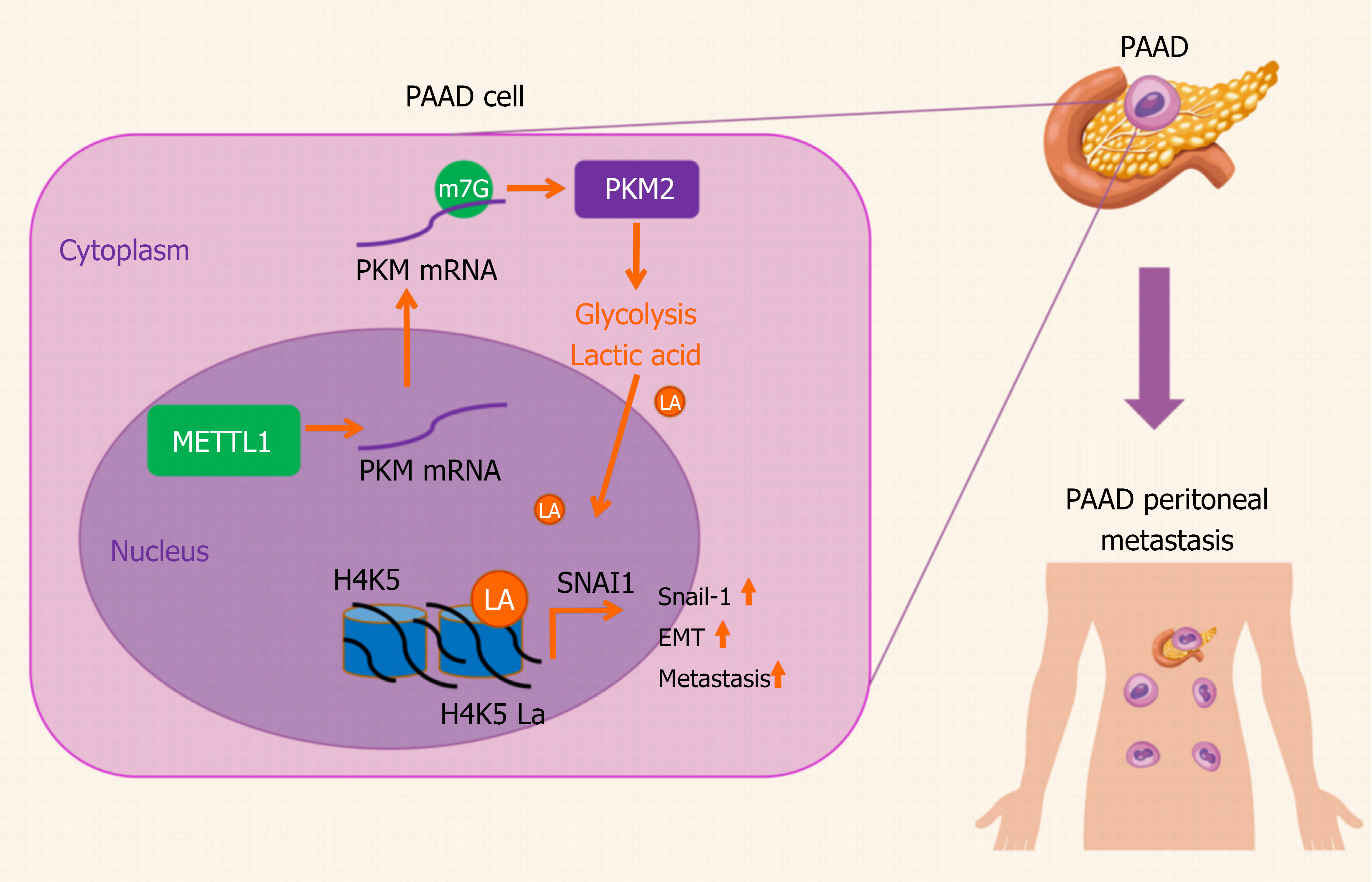
Figure 6 The mechanism of METTL1 promoting peritoneal metastasis of pancreatic adenocarcinoma via the N7-methylguanosine modi
- Citation: Yang C, Qiu CZ, Yin ZG, Mao Y, Ge XS, Wang F. METTL1-mediated N7-methylguanosine modification promotes glycolysis and H4K5 lactylation-dependent Snail-1 activation in pancreatic adenocarcinoma peritoneal metastasis. World J Gastroenterol 2027; 33(3): 123673
- URL: https://www.wjgnet.com/1007-9327/full/v33/i3/123673.htm
- DOI: https://dx.doi.org/10.3748/wjg.123673