
















Copyright: ©Author(s) 2026.
World J Gastroenterol. Dec 28, 2026; 32(48): 123390
Published online Dec 28, 2026. doi: 10.3748/wjg.123390
Published online Dec 28, 2026. doi: 10.3748/wjg.123390
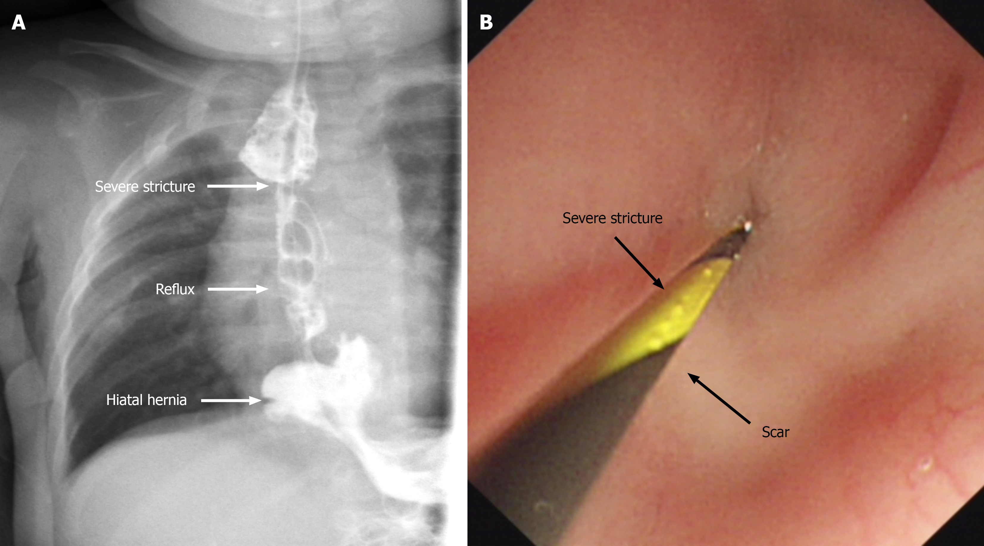
Figure 1 Preoperative imaging findings.
A: Upper gastrointestinal contrast study showing severe anastomotic stricture, hiatal hernia, and gastroesophageal reflux; B: Gastroscopy revealing a cicatricial stenotic orifice with a luminal diameter of approximately 1 mm.
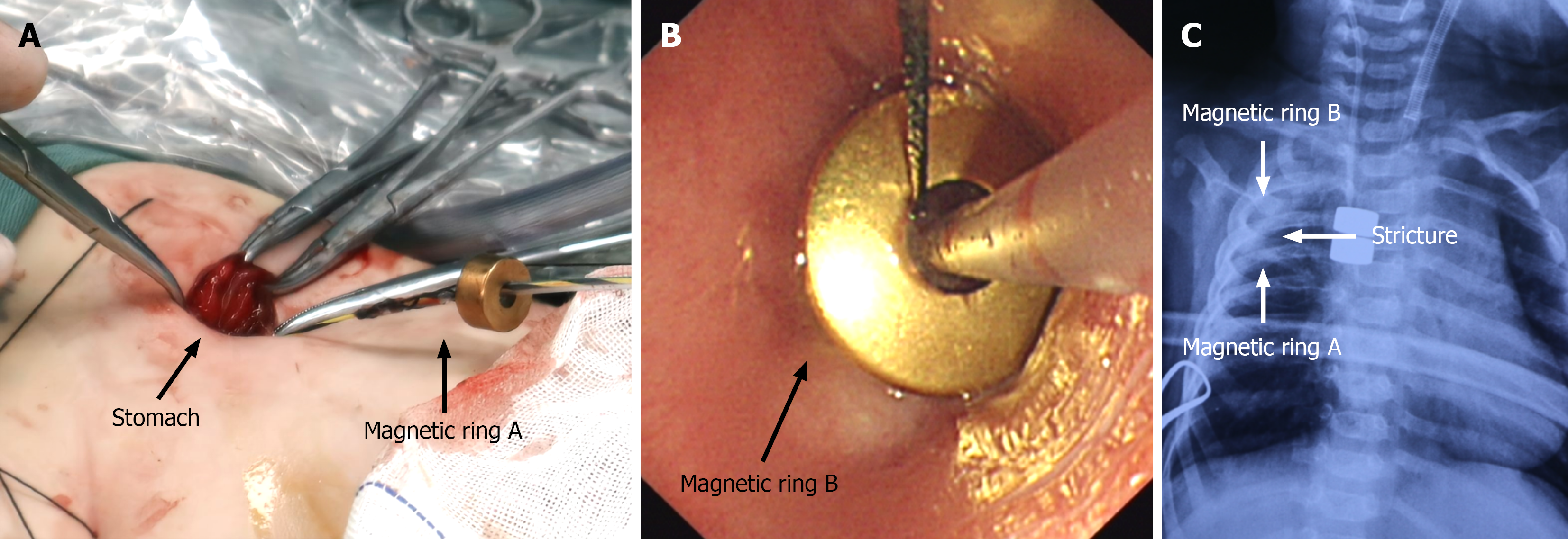
Figure 2 Intraoperative magnetic recanalization.
A: Magnetic ring A was delivered orally and positioned at the superior margin of the stricture under endoscopic guidance; B: Magnetic ring B was advanced retrogradely through a small anterior gastrotomy to the inferior margin of the stricture; C: Postoperative chest radiograph confirming proper alignment of the paired magnetic rings, hiatal hernia repair, and a smooth diaphragmatic contour.
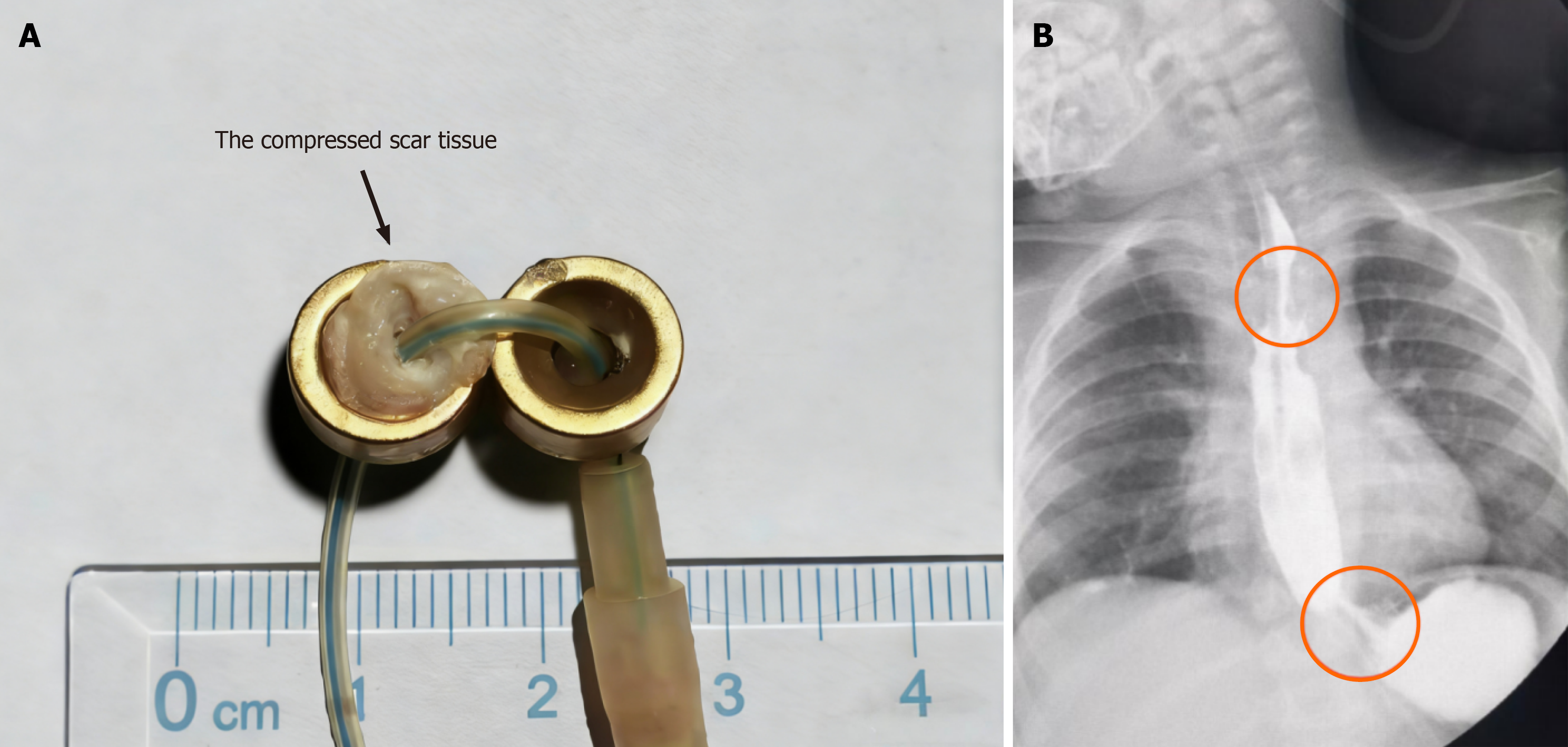
Figure 3 Postoperative magnet removal and outcome.
A: The magnetic rings were removed through the original gastrotomy at postoperative day 32, revealing complete excision of scar tissue within the crateriform depression; B: Upper gastrointestinal contrast study after magnet removal demonstrating restored esophageal patency (upper circle), with the gastric fundus situated below the diaphragm and resolution of gastroesophageal reflux (lower circle).
- Citation: Liu SQ, Li SQ, Sun YM, Li YF. Combined magnetic recanalization and laparoscopic antireflux surgery for complex esophageal comorbidities in an infant with congenital esophageal atresia. World J Gastroenterol 2026; 32(48): 123390
- URL: https://www.wjgnet.com/1007-9327/full/v32/i48/123390.htm
- DOI: https://dx.doi.org/10.3748/wjg.123390