
















Copyright: ©Author(s) 2026.
World J Gastroenterol. Nov 14, 2026; 32(42): 120674
Published online Nov 14, 2026. doi: 10.3748/wjg.120674
Published online Nov 14, 2026. doi: 10.3748/wjg.120674
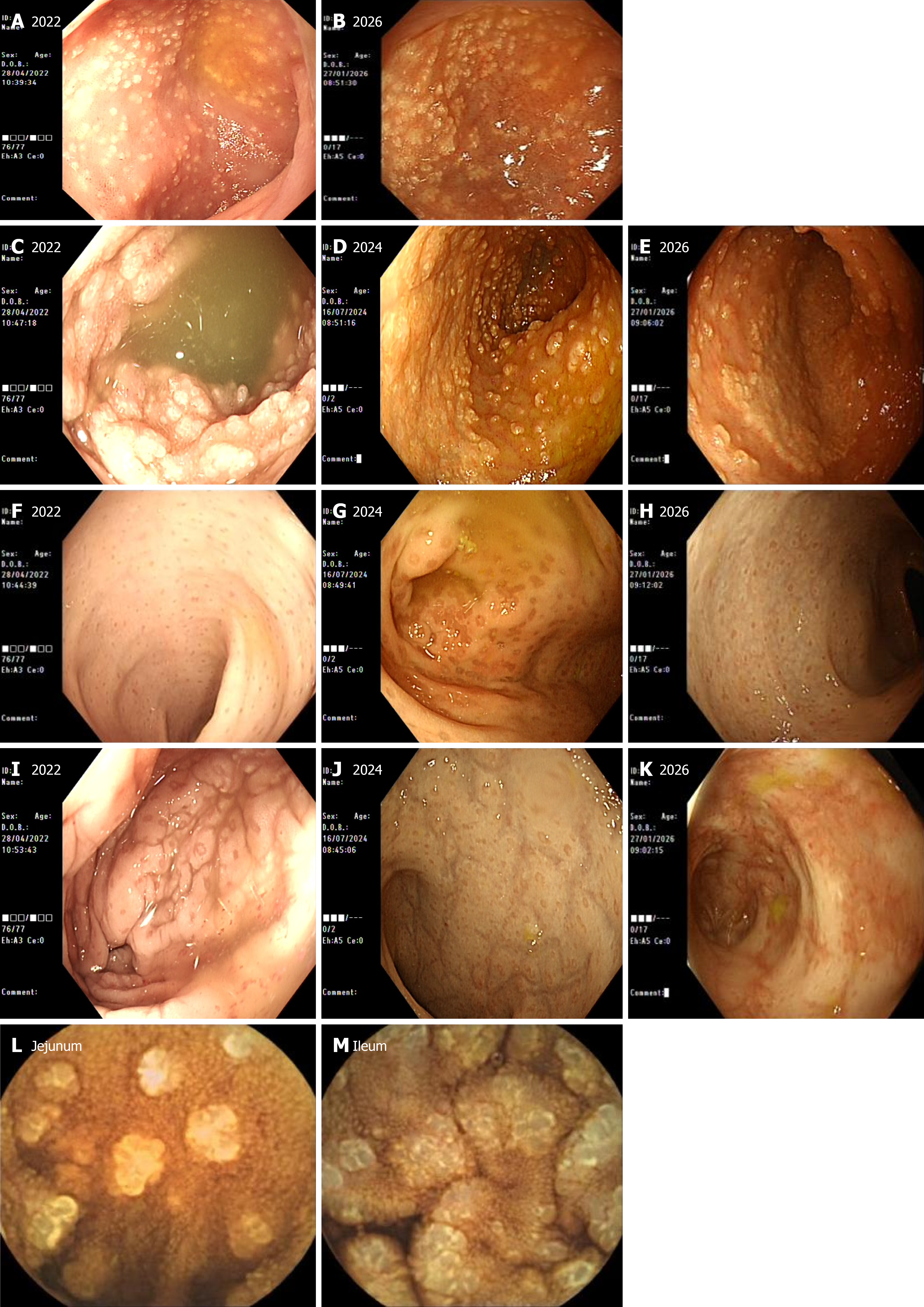
Figure 1 Endoscopic findings from 2022, 2024, and 2026 showing the duodenal bulb, terminal ileum, colon, jejunum and ileum.
A and B: Gastroscopy revealed diffuse yellowish-white granular protrusions in the duodenal bulb, resembling a tofu-like appearance in 2022 and 2026; C-E: Colonoscopy demonstrated extensive yellowish-white proliferative and elevated lesions in the mucosa of the terminal ileum, with variable lesion sizes and focal confluent patches in 2022, 2024, and 2026; F-H: The mucosa of the entire colon appeared pale, with numerous yellowish-brown spotted lesions observed in 2022, 2024, 2026; I-K: The mucosa of the rectum appeared yellowish-white with numerous yellowish-brown spotted lesions in 2022, 2024, and 2026; L and M: Capsule endoscopy revealed yellowish-white, dense, granular proliferative changes in portions of the jejunum and ileum, similar to the mucosal changes seen in the duodenal bulb in 2022.
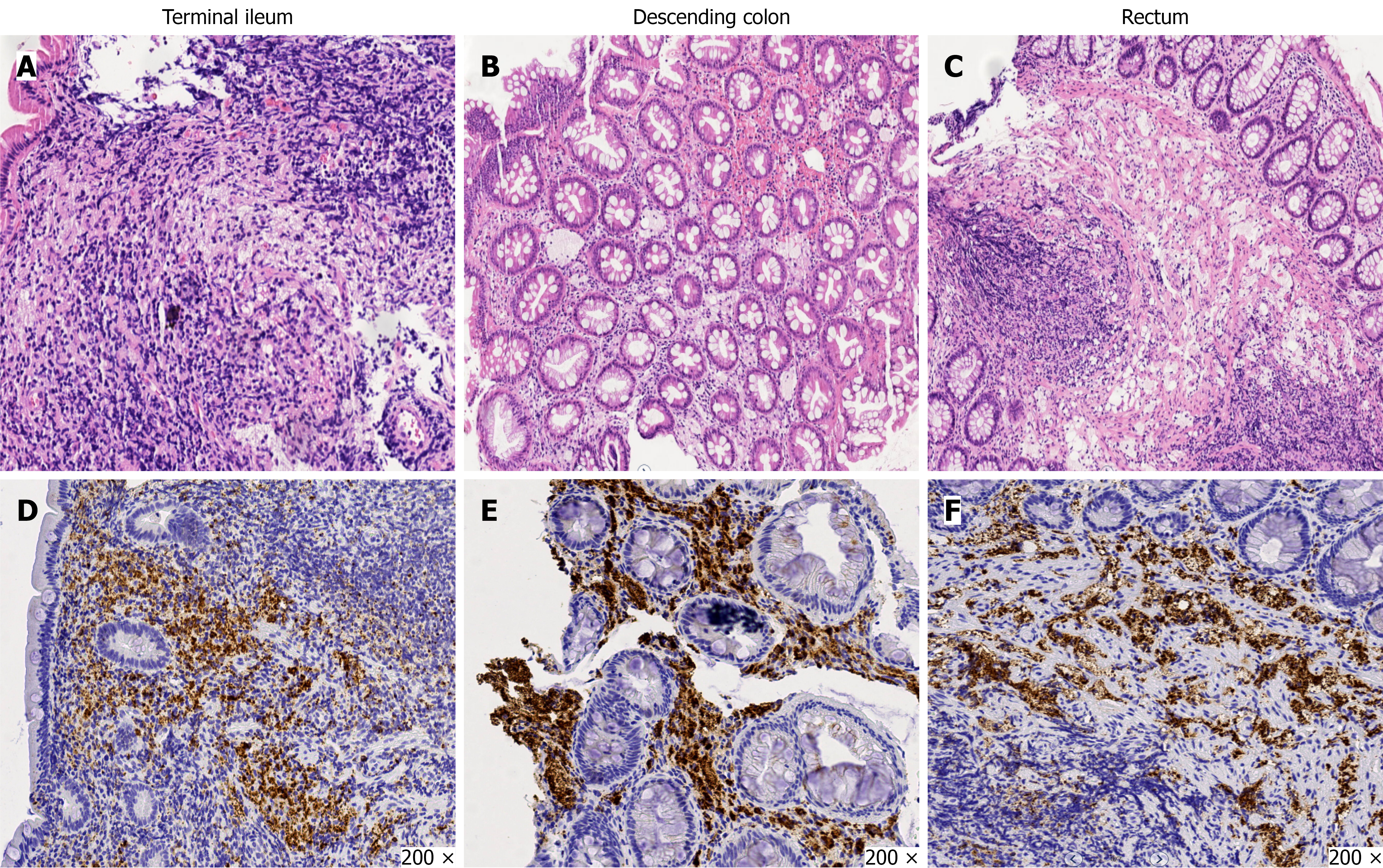
Figure 2 Pathology images.
A-C: Biopsy revealed chronic inflammation of the mucosa with lymphoid follicle infiltration in the lamina propria and submucosa, along with aggregation of foamy cells; D-F: Immunohistochemical staining revealed CD68 positivity, confirming a macrophage origin (× 200).
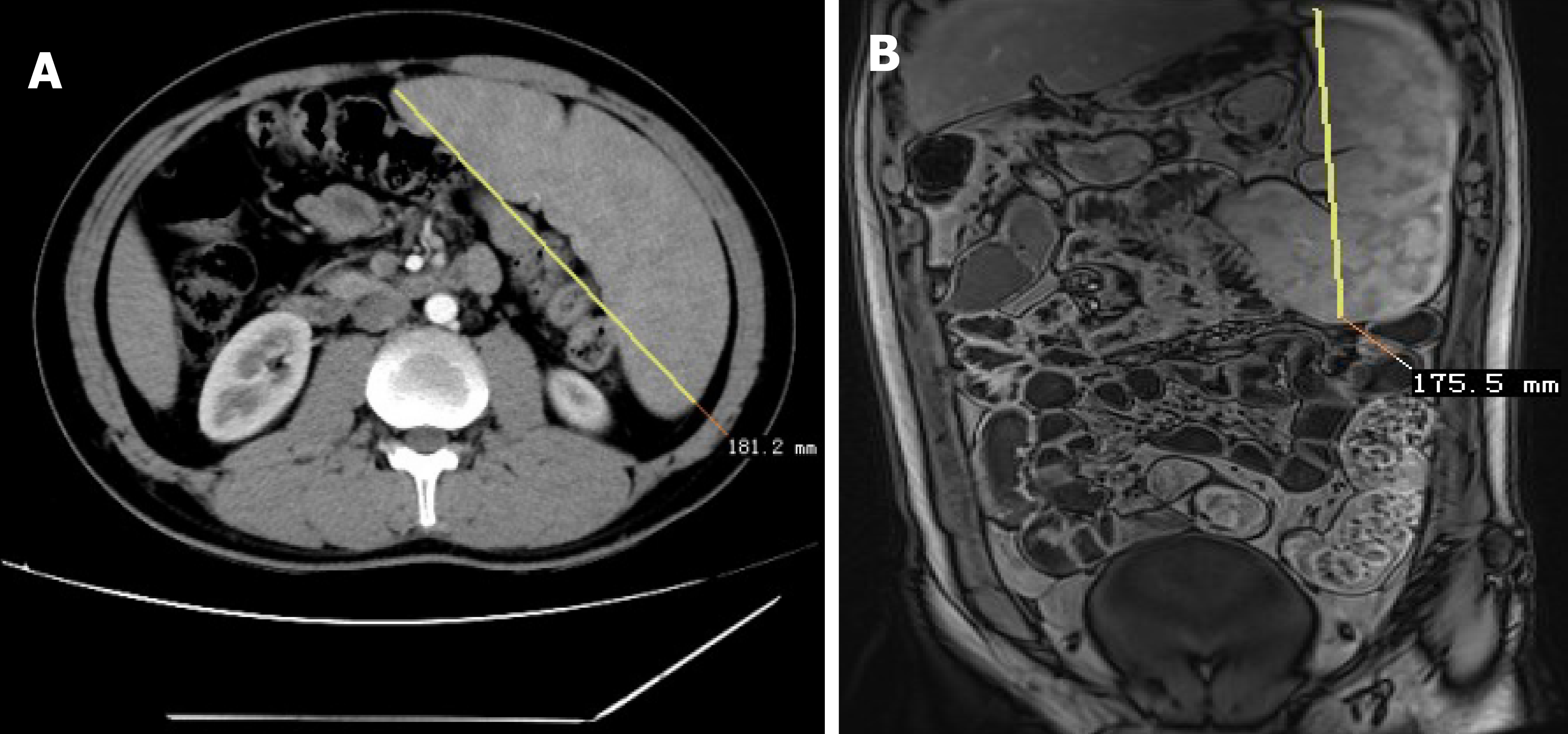
Figure 3 Abdominal computed tomography and small bowel magnetic resonance images.
A: Abdominal computed tomography demonstrated a splenic longitudinal diameter of approximately 18.1 cm on a specific imaging plane; B: The longest diameter of the spleen was 17.55 cm.
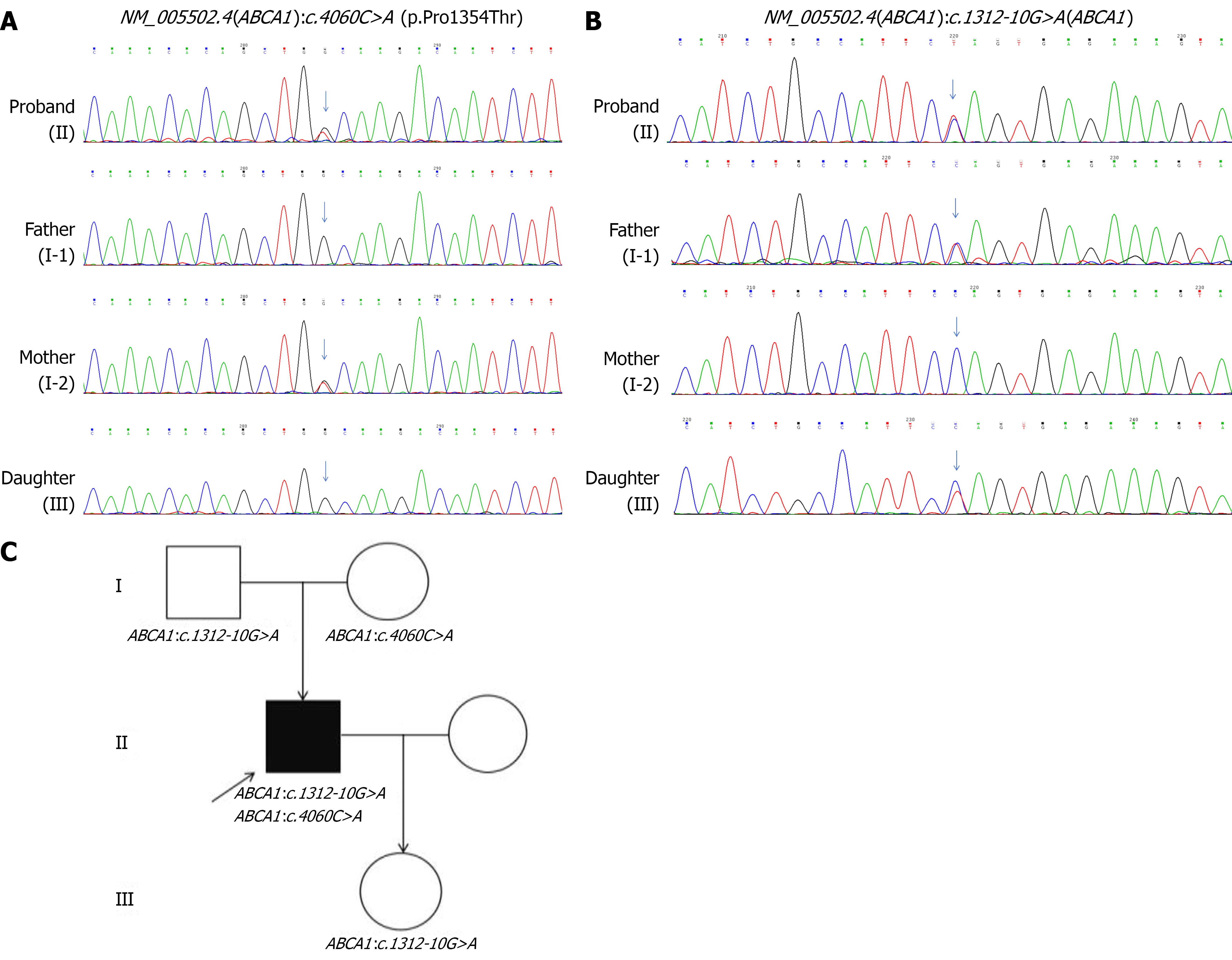
Figure 4 Genetic test results.
A and B: Sanger sequencing chromatogram, the patient’s mother carries the c.4060C>A:P.Pro1354Thr mutation, the patient’s father carries the c.1312-10G>A mutation, and the patient’s daughter also carries the ABCA1 (NM_005502.4):c.1312-10G>A; C: Pedigrees.
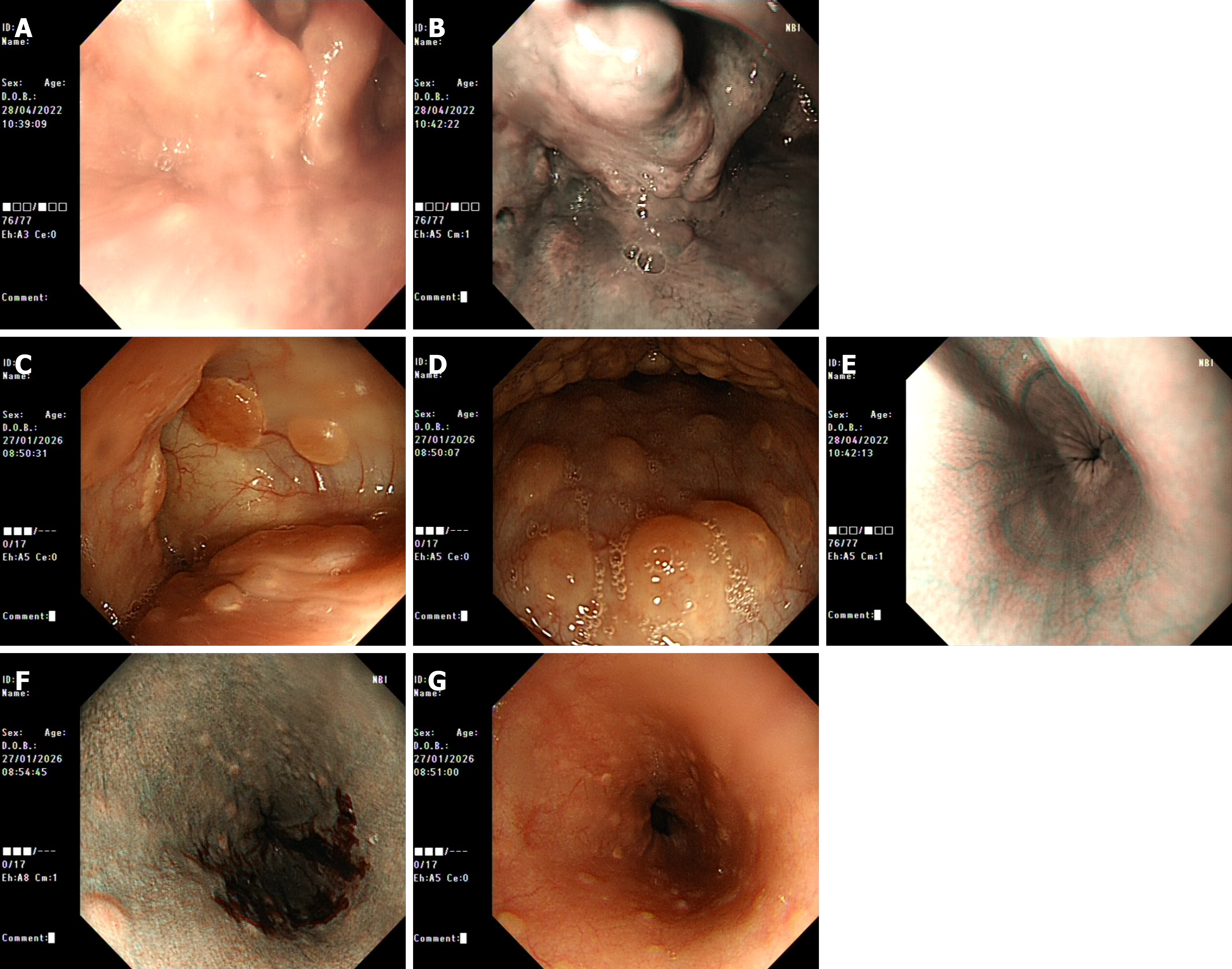
Figure 5 Comparison of local endoscopic findings between 2022 and 2026.
A and B: No significant abnormalities were observed in the oropharyngeal mucosa in 2022; C and D: Numerous round nodular elevated lesions with a yellowish surface were seen in the pharynx and tongue in 2026; E: No significant abnormalities were observed in the esophageal mucosa in 2022; F and G: Scattered granular hyperplasia with yellowish discoloration was noted in the lower esophagus in 2026.
- Citation: Xu XF, Wang JW, Wang LY, Chen CX. Tangier disease with heterozygous mutations in the ABCA1 gene: A case report. World J Gastroenterol 2026; 32(42): 120674
- URL: https://www.wjgnet.com/1007-9327/full/v32/i42/120674.htm
- DOI: https://dx.doi.org/10.3748/wjg.120674