
















Copyright: ©Author(s) 2026.
World J Gastroenterol. Nov 7, 2026; 32(41): 120464
Published online Nov 7, 2026. doi: 10.3748/wjg.120464
Published online Nov 7, 2026. doi: 10.3748/wjg.120464
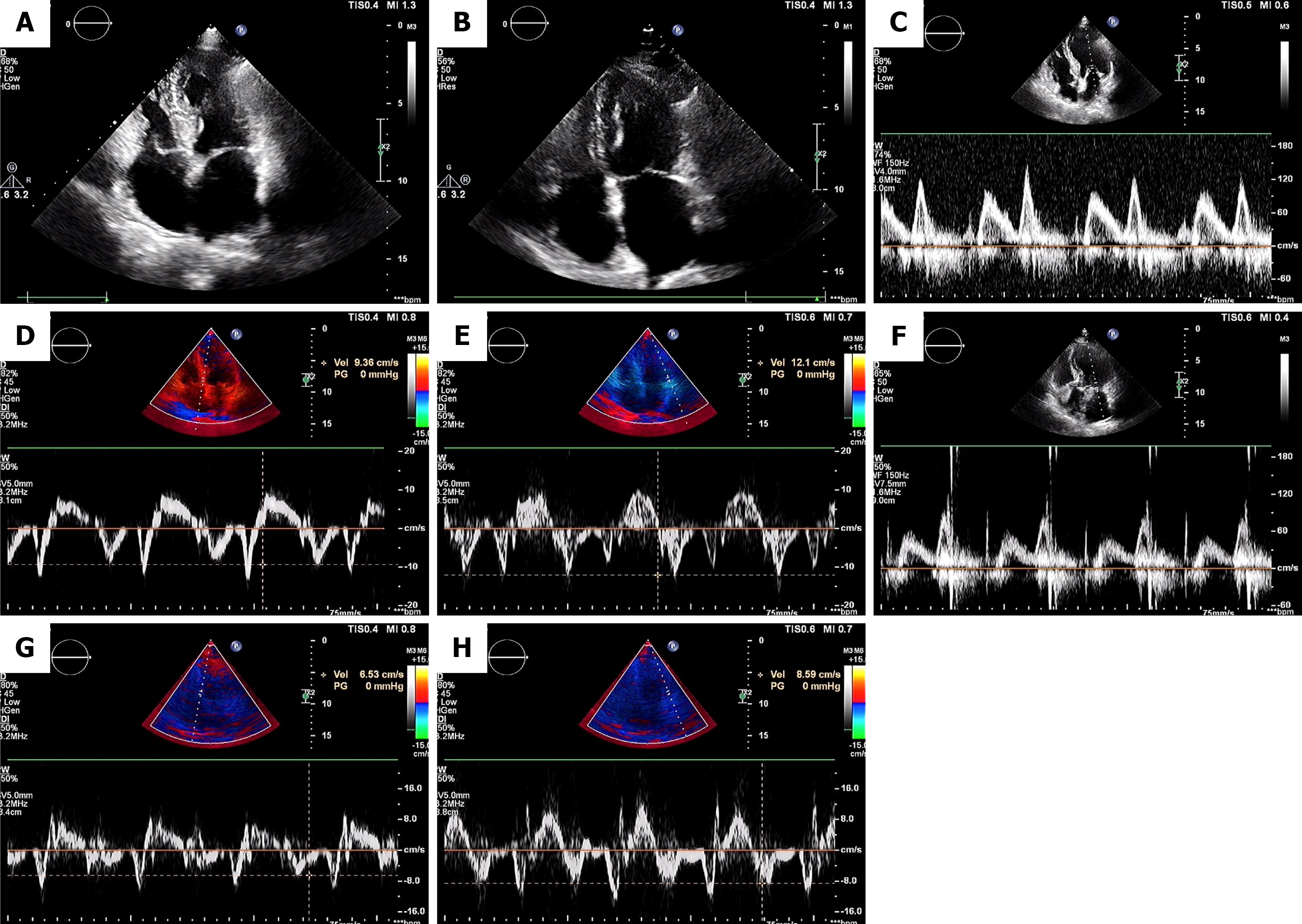
Figure 1 Echocardiographic assessment of left atrial size and left ventricular diastolic function.
A and B: Apical four-chamber views showing left atrial size. Normal left atrial volume index (≤ 34 mL/m2) (A). Increased left atrial volume index (> 34 mL/m2) indicating left atrial enlargement (B); C-H: Parameters for diastolic function assessment. Mitral inflow pulse-wave Doppler spectra (E and A waves) in two patients (C, F). Septal (D, G) and lateral (E, H) early diastolic mitral annular velocities (e’) obtained via tissue Doppler imaging from an apical four-chamber view, displaying normal (septal e’ ≥ 7 cm/second, lateral e’ ≥ 10 cm/second; D, E) and reduced (septal e’ < 7 cm/second, lateral e’ < 10 cm/second; G, H) e’ velocities. Average E/e’ ratio ≤ 14 indicates normal filling pressure; > 14 suggests elevated filling pressure.
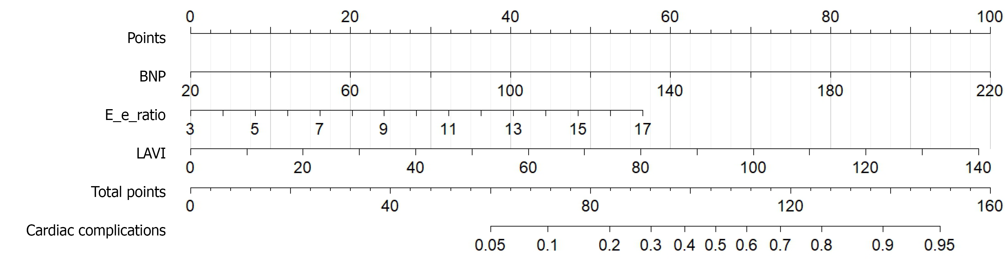
Figure 2 Nomogram for cardiac complications within 30 days postoperatively.
BNP: B-type natriuretic peptide; LAVI: Left atrial volume index.
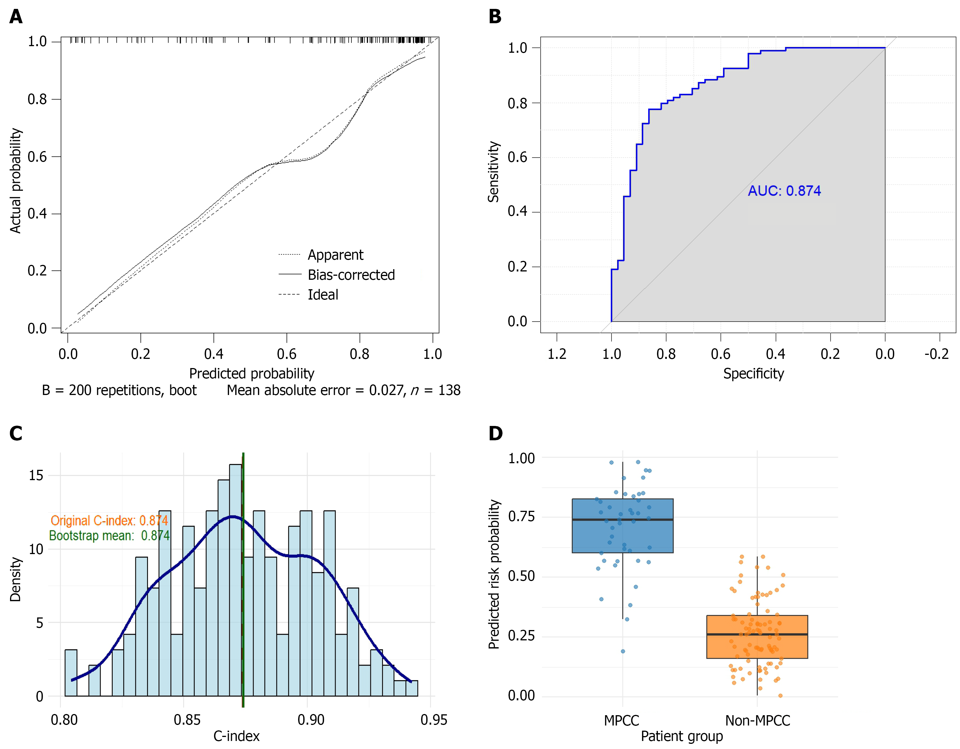
Figure 3 Development and internal validation of the nomogram for predicting major postoperative cardiac complications.
A: Calibration curve; B: Receiver operating characteristic curve; C: Distribution of the C-index from bootstrap internal validation; D: Distribution of predicted probabilities. AUC: Area under the receiver operating characteristic curve; MPCC: Major postoperative cardiac complication.
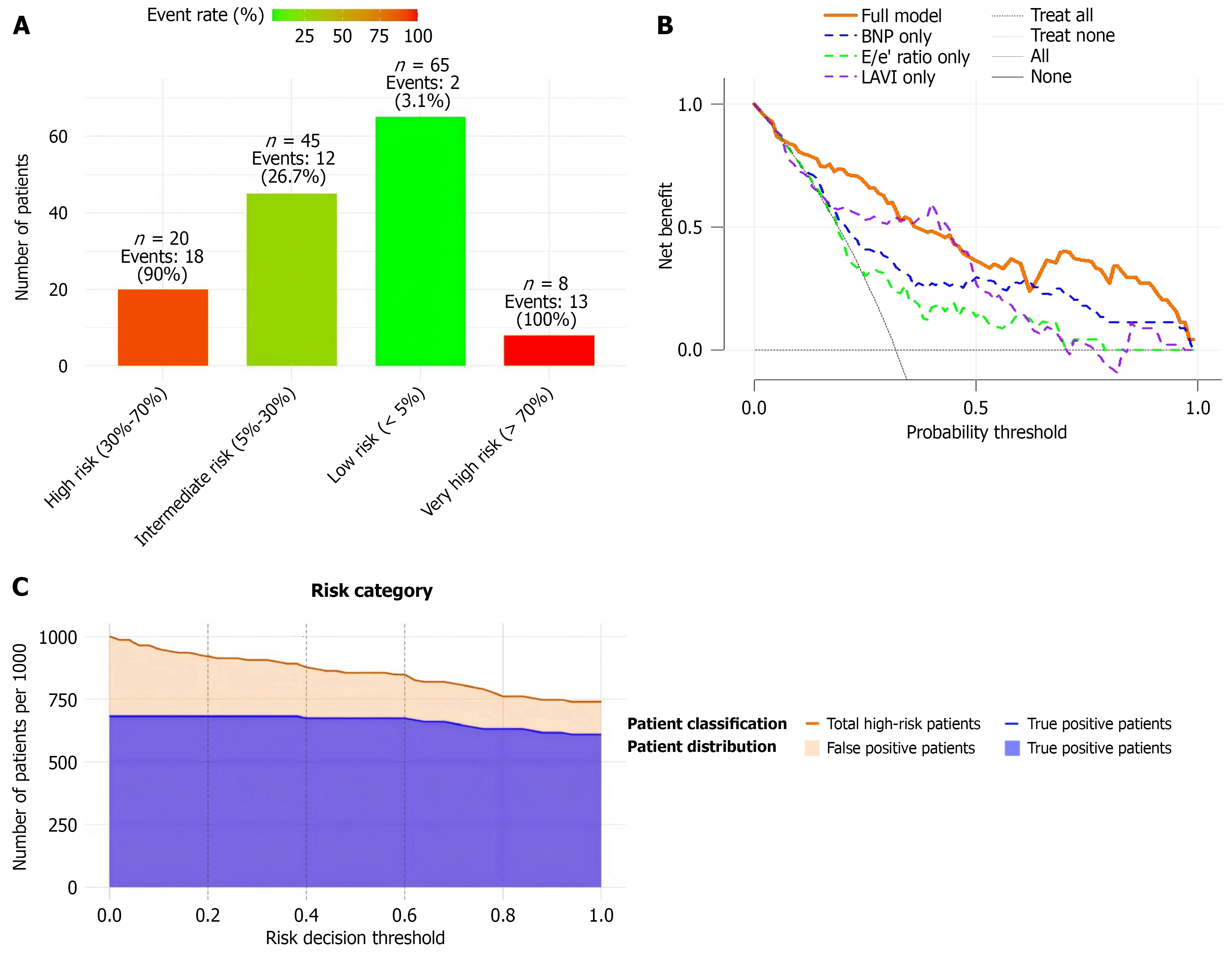
Figure 4 Clinical validation of the prediction nomogram.
A: Nomogram-based risk stratification: B: Decision curve analysis; C: Clinical impact curve. BNP: B-type natriuretic peptide; LAVI: Left atrial volume index.
- Citation: Jin S, Wang FF, Lyu SC, Zhao X, He Q, Lyu XZ. Preoperative echocardiography-based nomogram for postoperative cardiac complications in pancreatic cancer with vascular invasion. World J Gastroenterol 2026; 32(41): 120464
- URL: https://www.wjgnet.com/1007-9327/full/v32/i41/120464.htm
- DOI: https://dx.doi.org/10.3748/wjg.120464