











BPG is committed to discovery and dissemination of knowledge
Editorial
Copyright: ©Author(s) 2026.
World J Gastroenterol. Oct 28, 2026; 32(40): 121301
Published online Oct 28, 2026. doi: 10.3748/wjg.121301
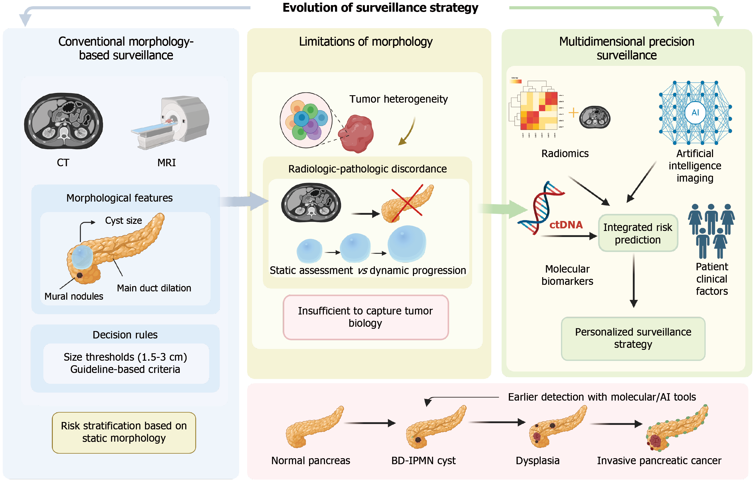
Figure 1
Figure 1 Conceptual evolution from morphology-based surveillance to multidimensional precision surveillance in branch-duct intraductal papillary mucinous neoplasms. The figure illustrates a stepwise transition from conventional morphology-based assessment to an integrated, precision surveillance framework. In the left panel, conventional surveillance relies on cross-sectional imaging modalities, including computed tomography and magnetic resonance imaging, where risk stratification is primarily based on static morphological features such as cyst size, main pancreatic duct dilation, and mural nodules. These features are typically interpreted using predefined threshold criteria (e.g., 1.5 cm to 3 cm), with the accumulation of multiple worrisome features associated with increased malignancy risk. However, as shown in the middle panel, this morphology-based approach has important limitations. Tumor biological heterogeneity cannot be adequately captured by imaging alone, leading to radiologic-pathologic discordance. In addition, reliance on static snapshots fails to reflect the dynamic nature of tumor evolution over time. Collectively, these limitations restrict accurate characterization of tumor biology and risk. In contrast, the right panel presents a multidimensional precision surveillance paradigm that integrates diverse data sources, including radiomics, molecular biomarkers (e.g., circulating tumor DNA), artificial intelligence-based imaging analysis, and patient-specific clinical factors. Through multimodal data integration, this approach enables more robust risk prediction and supports individualized surveillance and management strategies. The bottom schematic depicts the continuum of disease progression from normal pancreas to branch-duct intraductal papillary mucinous neoplasms, dysplasia, and invasive pancreatic cancer. It highlights how the incorporation of molecular and artificial intelligence-assisted tools may facilitate earlier detection and intervention within this progression spectrum. CT: Computed tomography; MRI: Magnetic resonance imaging; ctDNA: Circulating tumor DNA; AI: Artificial intelligence; BD-IPMN: Branch-duct intraductal papillary mucinous neoplasm.


Write to the Help Desk