Copyright
©The Author(s) 2024.
World J Gastroenterol. Mar 7, 2024; 30(9): 994-998
Published online Mar 7, 2024. doi: 10.3748/wjg.v30.i9.994
Published online Mar 7, 2024. doi: 10.3748/wjg.v30.i9.994
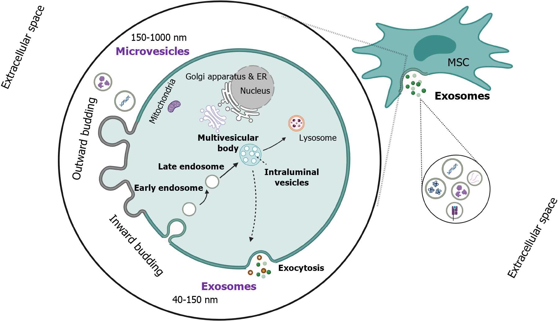
Figure 1 Biogenesis of extracellular vesicles.
Exosome generation is initiated by the budding of the inward plasma membrane and the internalization of transmembrane proteins, forming early endosomes, which are further maturated into late endosomes. Under the contribution of endosomal sorting complexes 0-III, the invagination of the late endosomal membrane forms the intraluminal vesicles (ILVs). Eventually, multivesicular bodies (MVBs), which have several ILVs in their lumen, are formed. Another site of cargo, besides the membrane, is the trans-Golgi complex or cytoplasm. Afterward, MVBs are either fused with cell membrane for the exocytosis of exosomes or degraded in lysosomes. However, they can form amphisomes via fusion with autophagosomes, which are either degraded in lysosomes or fuse with plasma membrane to release the vesicles into the extracellular space. A broad spectrum of cells/tissues, including mesenchymal stem cells, can produce extracellular vesicles. ILV: Intraluminal vesicles; MSC: Mesenchymal stem cells. Created with “BioRender.com” (Supplementary material)[20].
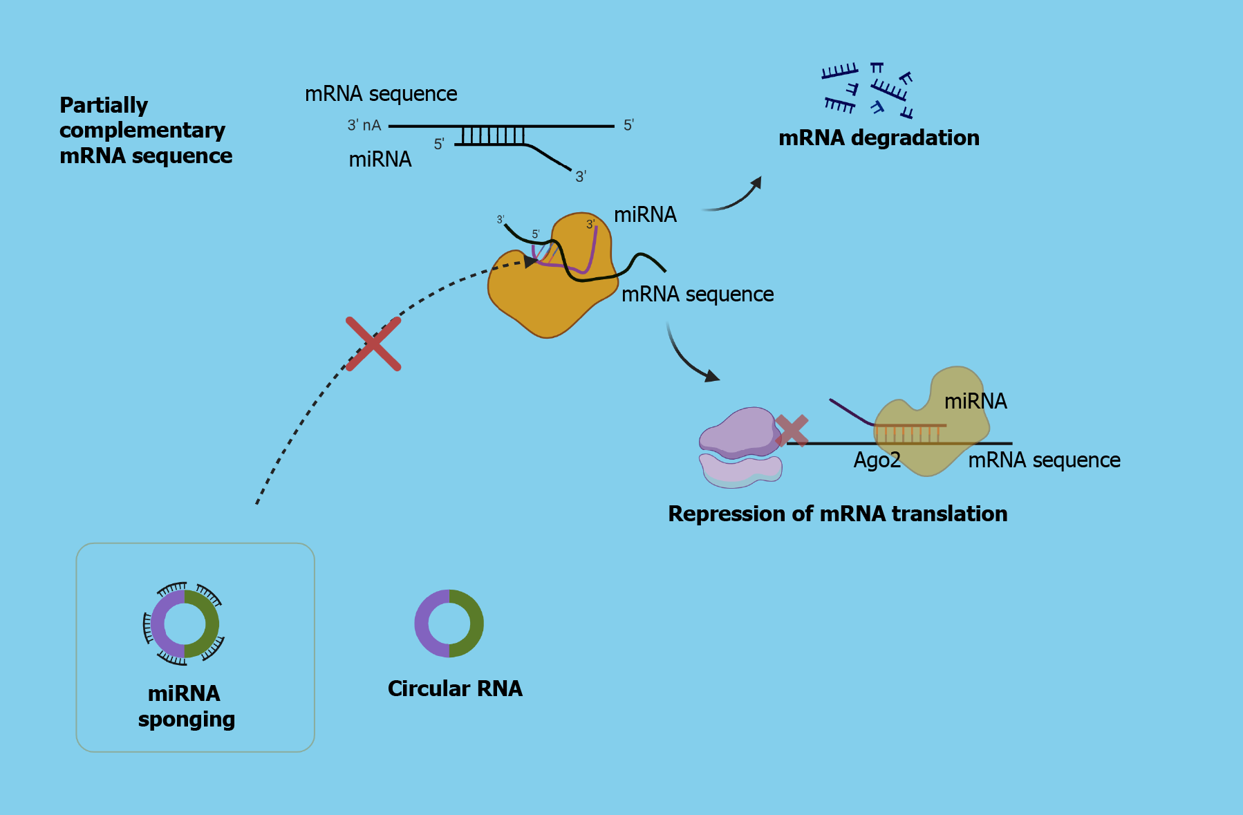
Figure 2 Role of circular RNA as “microRNA sponge”.
After the active microRNA (miRNA) strand (leading strand/mature miRNA) is loaded on the RISC complex, the so-called miRISC (seed sequence), which is capable of binding on a target mRNA strand, leads in the suppression of mRNA translation, silencing, or even degradation. Circular RNAs (circRNAs) are non-coding RNA molecules that have a pivotal contribution to gene expression, as they can protect mRNA translation from miRNAs, via miRNA sponging, interact with several proteins, including RNA-binding proteins and polymerase II, and translocate them. circRNA: Circular RNAs; mRNA: Messenger RNA; miRs: MicroRNAs. Created with “BioRender.com” (Supplementary material)[20].
- Citation: Papadopoulos N, Trifylli EM. Role of exosomal circular RNAs as microRNA sponges and potential targeting for suppressing hepatocellular carcinoma growth and progression. World J Gastroenterol 2024; 30(9): 994-998
- URL: https://www.wjgnet.com/1007-9327/full/v30/i9/994.htm
- DOI: https://dx.doi.org/10.3748/wjg.v30.i9.994


