Copyright
©The Author(s) 2024.
World J Gastroenterol. Mar 7, 2024; 30(9): 1132-1142
Published online Mar 7, 2024. doi: 10.3748/wjg.v30.i9.1132
Published online Mar 7, 2024. doi: 10.3748/wjg.v30.i9.1132
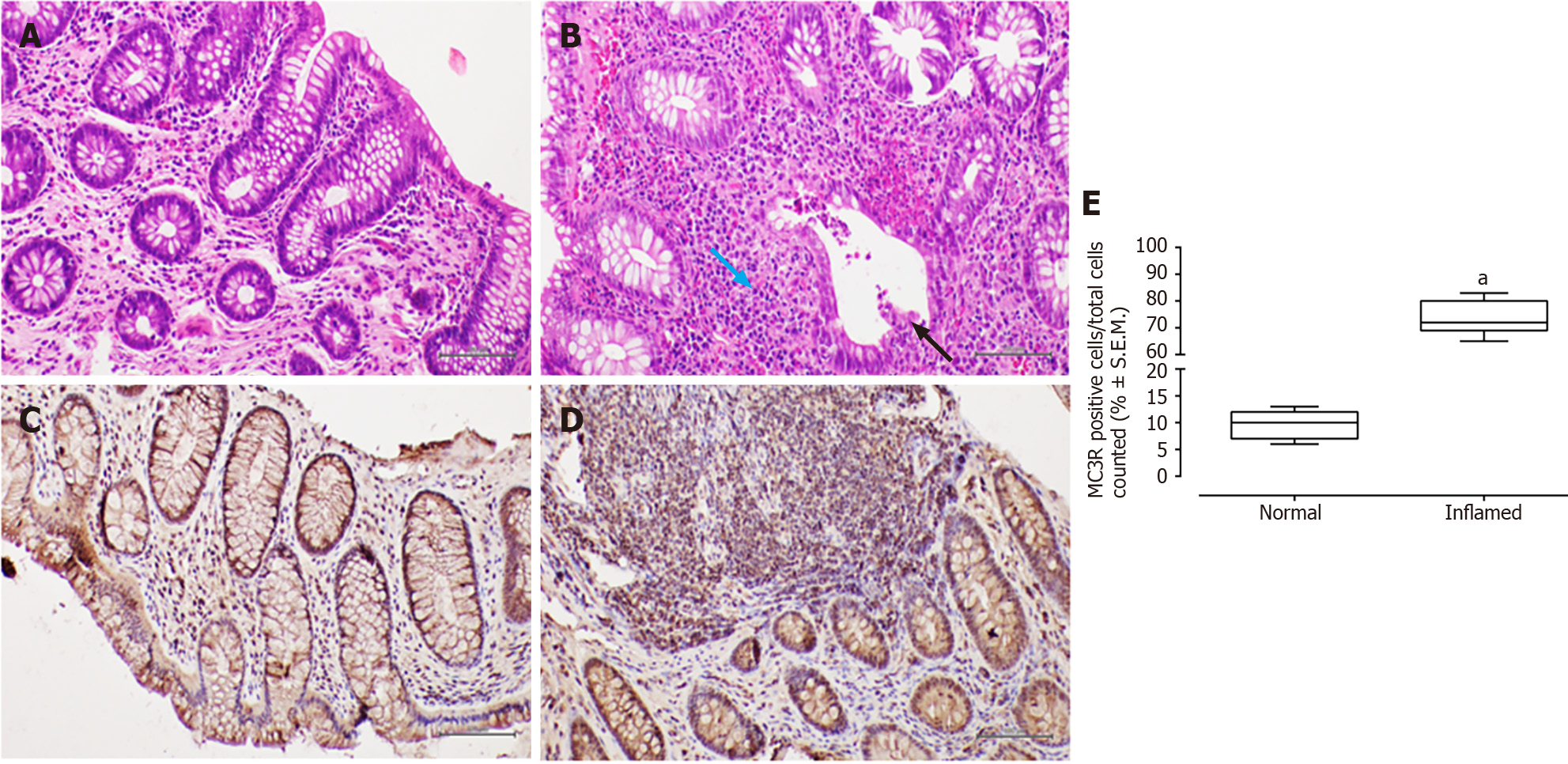
Figure 1 Melanocortin 3 receptor expression in samples from individuals with Crohn’s disease.
A and B: Representative histological sections stained with haematoxylin-eosin for normal (A) and inflamed (B) mucosa. The blue arrow highlights the dense chronic inflammatory infiltrate, while the black arrow indicates histological activity with cryptitis and microabscesses; C and D: Immunohistochemical staining in the former (C) and latter (D) is also depicted. The rate of Melanocortin 3 receptor (MC3R) cell positivity, as determined by immunohistochemistry, was significantly higher in the inflamed mucosa compared to the normal mucosa; E: The data are presented as % ± SEM of MC3R positive cells relative to the total cells counted. Scale bar = 500 µm. aP < 0.01.
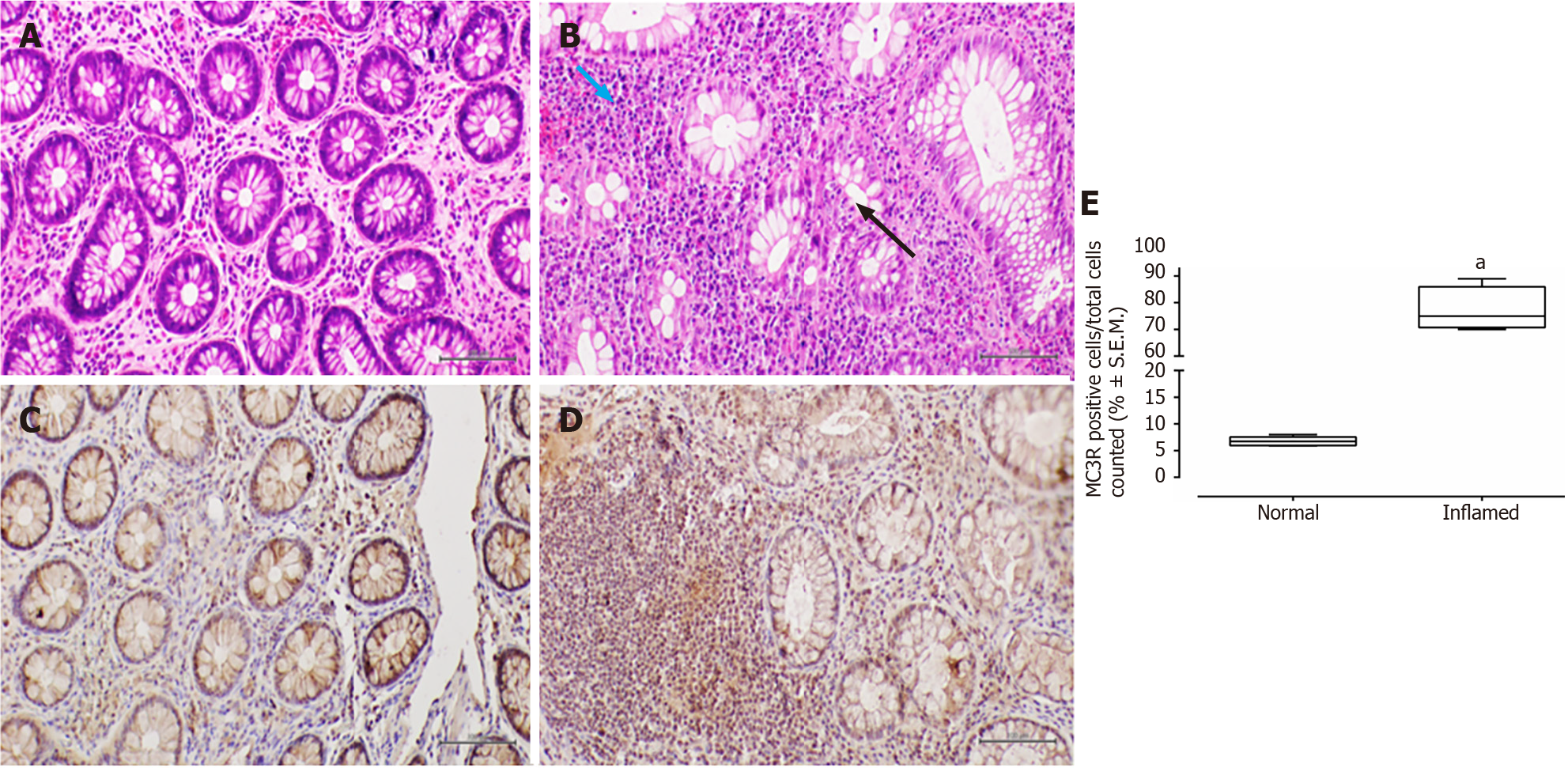
Figure 2 Melanocortin 3 receptor expression in samples from individuals with ulcerative colitis.
A and B: Representative histological sections stained with haematoxylin-eosin for normal (A) and inflamed (B) mucosa. The blue arrow highlights the dense chronic inflammatory infiltrate, while the black arrow indicates histological activity with cryptitis; C and D: Immunohistochemical staining in the former (C) and latter (D) is also presented. The rate of Melanocortin 3 receptor (MC3R) cell positivity, determined through immunohistochemistry, was significantly higher in the inflamed mucosa compared to the normal mucosa; E: The data are expressed as % ± SEM of MC3R positive cells relative to the total cells counted. Scale bar = 500 µm. aP < 0.01.
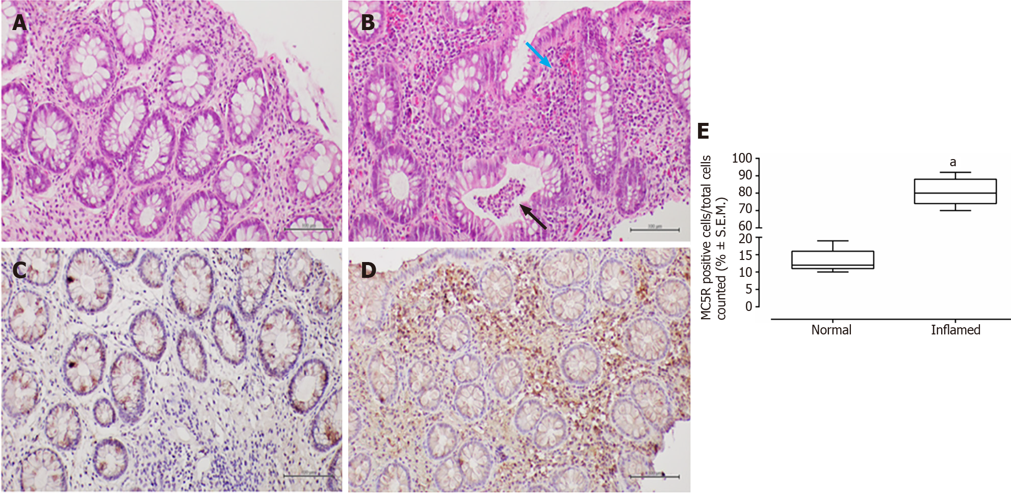
Figure 3 Melanocortin 5 receptor expression in samples from individuals with Crohn's disease.
A and B: Representative histological sections stained with haematoxylin-eosin for normal (A) and inflamed (B) mucosa. The blue arrow highlights the dense chronic inflammatory infiltrate, while the black arrow indicates histologic activity with microabscesses; C and D: Immunohistochemical staining in the former (C) and latter (D) is also presented. The rate of Melanocortin 5 receptor (MC5R) cell positivity, determined through immunohistochemistry, was significantly higher in the inflamed mucosa compared to the normal mucosa; E: The data are expressed as % ± SEM of MC5R positive cells relative to the total cells counted. Scale bar = 500 µm. aP < 0.01.
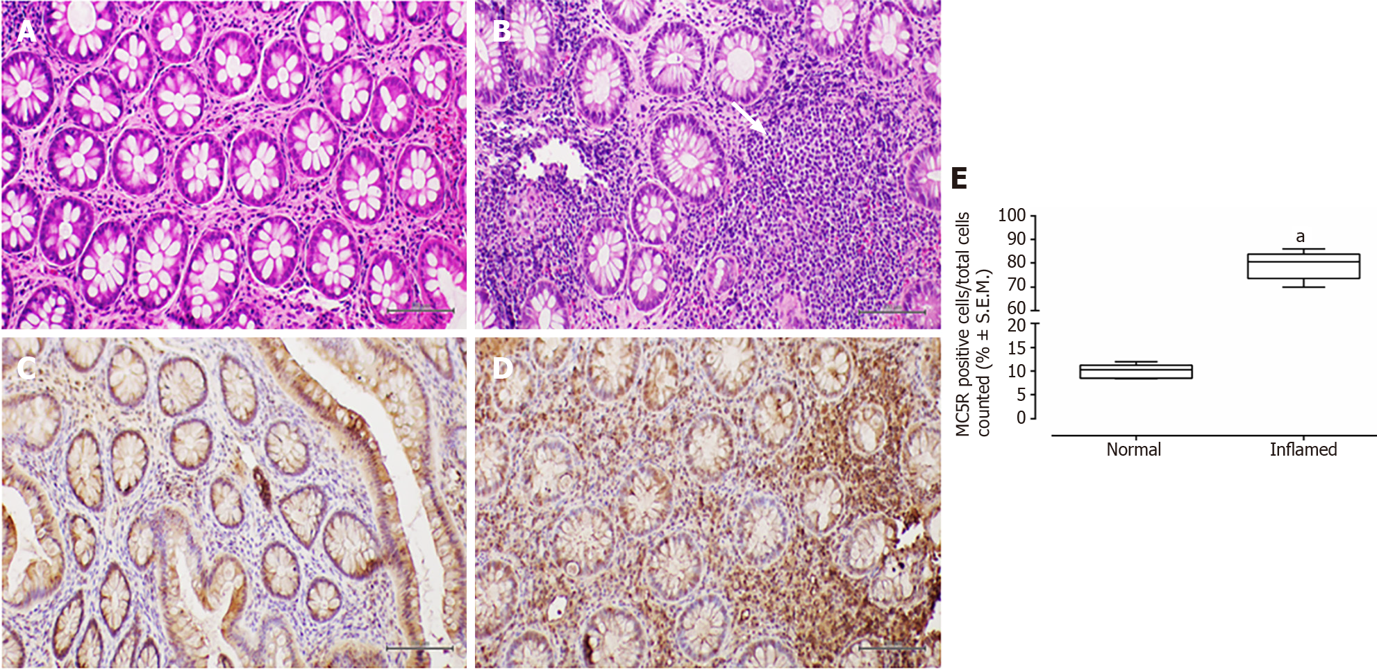
Figure 4 Melanocortin 5 receptor expression in samples from individuals with ulcerative colitis.
A and B: Representative histological sections stained with haematoxylin-eosin for normal (A) and inflamed (B) mucosa. The white arrow highlights the dense chronic inflammatory infiltrate in inflamed mucosa; C and D: Immunohistochemical staining in the former (C) and latter (D) is also presented. The rate of Melanocortin 5 receptor (MC5R) cell positivity, determined through immunohistochemistry, was significantly higher in the inflamed mucosa compared to the normal mucosa; E: The data are expressed as % ± SEM of MC5R positive cells relative to the total cells counted. Scale bar = 500 µm. aP < 0.01.
- Citation: Gravina AG, Panarese I, Trotta MC, D'Amico M, Pellegrino R, Ferraraccio F, Galdiero M, Alfano R, Grieco P, Federico A. Melanocortin 3,5 receptors immunohistochemical expression in colonic mucosa of inflammatory bowel disease patients: A matter of disease activity? World J Gastroenterol 2024; 30(9): 1132-1142
- URL: https://www.wjgnet.com/1007-9327/full/v30/i9/1132.htm
- DOI: https://dx.doi.org/10.3748/wjg.v30.i9.1132


