Copyright
©The Author(s) 2024.
World J Gastroenterol. Feb 21, 2024; 30(7): 614-623
Published online Feb 21, 2024. doi: 10.3748/wjg.v30.i7.614
Published online Feb 21, 2024. doi: 10.3748/wjg.v30.i7.614
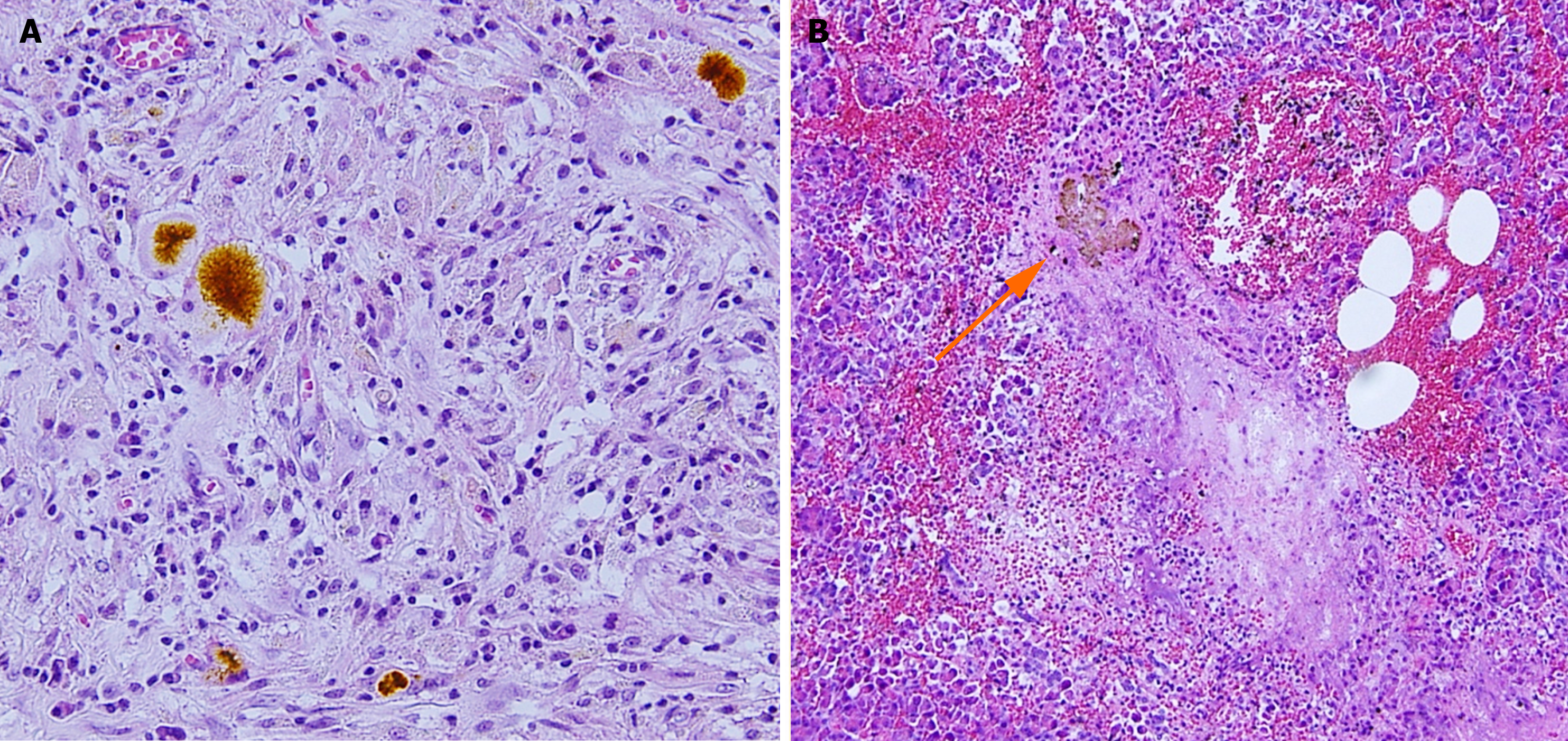
Figure 1 Histological evidence of bile reflux into the pancreas in a patient with gallstone necrotizing pancreatitis.
A: Bile pigments among the inflammatory granulomatous tissue; B: Concretion of bile pigment-like material (arrow), periductal necrosis with neutrophil infiltration, and bleeding. Citation: Isogai M, Kaneoka Y, Iwata Y. Histological evidence of bile reflux in necrotizing pancreatitis: A case report. Med Case Rep Study Protoc 2020; 1: e0016. Copyright ©The Author 2020. Published by Wolters Kluwer Health Inc[50].
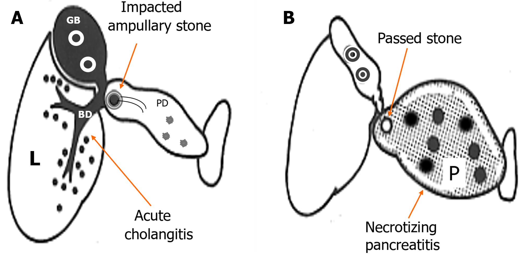
Figure 2 Subdivision of severe gallstone pancreatitis into gallstone cholangiopancreatitis and gallstone necrotizing pancreatitis.
A: Gallstone cholangiopancreatitis with persistent ampullary stone impaction and ascending acute cholangitis complicated with minimal or mild pancreatic inflammation caused by biliopancreatic obstruction. B: Gallstone necrotizing pancreatitis caused by the reflux of bile or possible duodenal contents into the pancreas, not complicated by acute biliary tract disease caused by the passage of stones. L: Liver; BD: Bile duct; GB: Gallbladder; PD: Pancreatic duct; P: Pancreas. Citation: Isogai M. Proposal of the term “gallstone cholangiopancreatitis” to specify gallstone pancreatitis that needs urgent endoscopic retrograde cholangiopancreatography. World J Gastrointest Endosc 2021; 13: 451-459. Copyright ©The Author(s) 2021. Published by Baishideng Publishing Group Inc[74].
- Citation: Isogai M. Pathophysiology of severe gallstone pancreatitis: A new paradigm. World J Gastroenterol 2024; 30(7): 614-623
- URL: https://www.wjgnet.com/1007-9327/full/v30/i7/614.htm
- DOI: https://dx.doi.org/10.3748/wjg.v30.i7.614


