Copyright
©The Author(s) 2023.
World J Gastroenterol. Oct 28, 2023; 29(40): 5557-5565
Published online Oct 28, 2023. doi: 10.3748/wjg.v29.i40.5557
Published online Oct 28, 2023. doi: 10.3748/wjg.v29.i40.5557
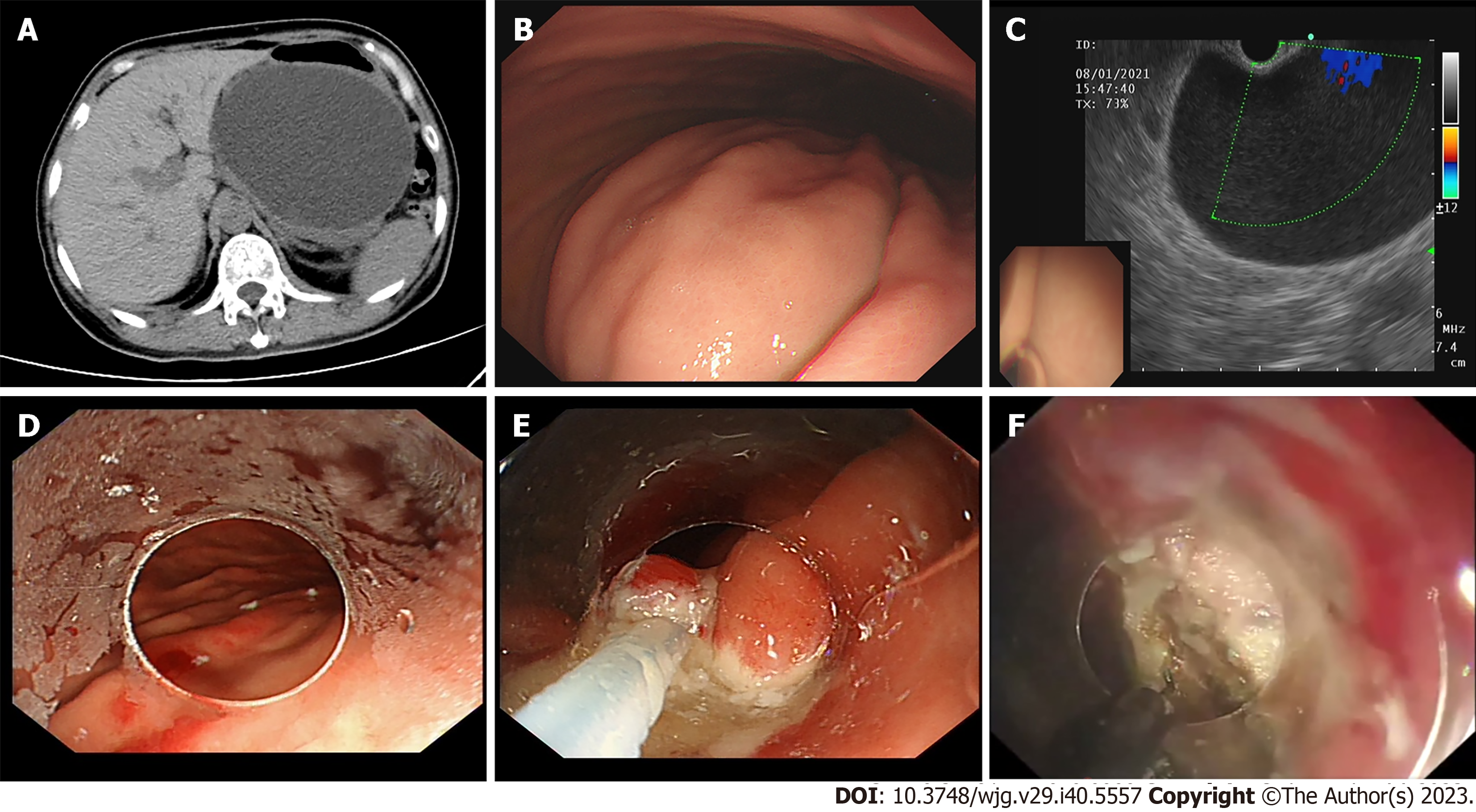
Figure 1 Endoscopic transgastric fenestration: From location to endoscopic full-thickness resection.
A: Preoperative computed tomography scan of (peri) pancreatic collections; B: Gastric bulge caused by (peri) pancreatic collections under upper endoscope view; C: Assessment of fenestration site under endoscopic ultrasound; D: Marked the mucous layer; E: Resect the mucosal layer by endoscopic mucosal resection; F: Full-thickness incision was operated.
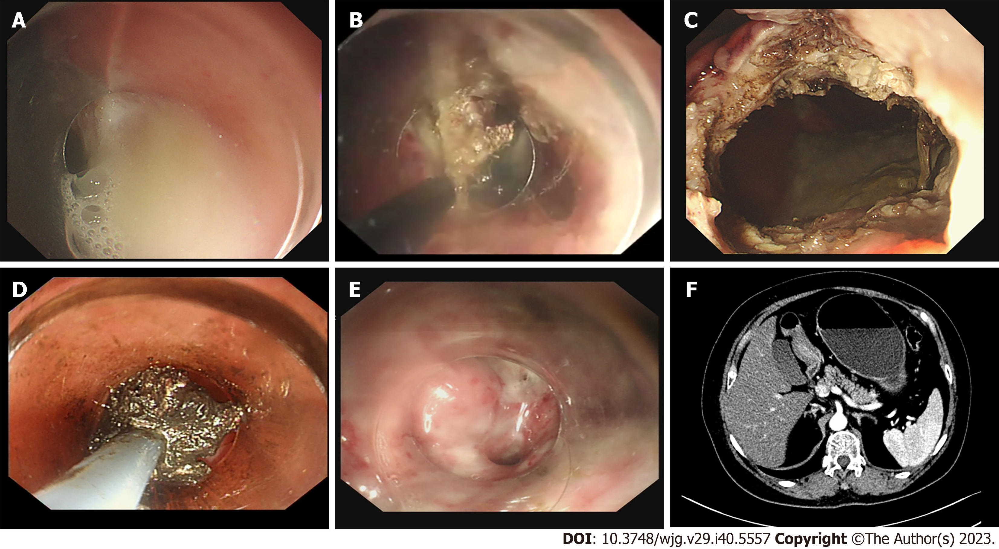
Figure 2 Endoscopic transgastric fenestration: Enlarge the aperture of opening and debridement.
A: Collections inflowing via the artificial fistula; B: Enlarged the aperture of opening with hook knife; C: The artificial fistula (about 2 cm in diameter) between the gastric wall and the cavity was made; D: Debridement of necrotic tissue under endoscopic direct vision; E: Endoscopic review of the fistula showed fistula almost healed one months later; F: The reviewed computed tomography scan one month after endoscopic transgastric fenestration.
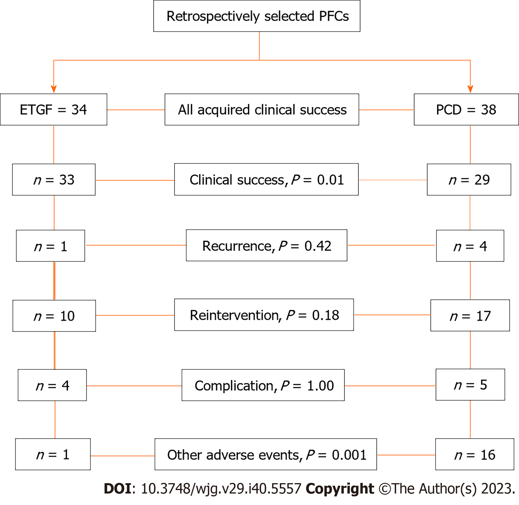
Figure 3 Flow-chart for this retrospective study.
ETGF: Endoscopic transgastric fenestration; PFCs: (peri) Pancreatic collections; PCD: Percutaneous drainage.
- Citation: Zhang HM, Ke HT, Ahmed MR, Li YJ, Nabi G, Li MH, Zhang JY, Liu D, Zhao LX, Liu BR. Endoscopic transgastric fenestration versus percutaneous drainage for management of (peri)pancreatic fluid collections adjacent to gastric wall (with video). World J Gastroenterol 2023; 29(40): 5557-5565
- URL: https://www.wjgnet.com/1007-9327/full/v29/i40/5557.htm
- DOI: https://dx.doi.org/10.3748/wjg.v29.i40.5557


