Copyright
©The Author(s) 2023.
World J Gastroenterol. Jan 21, 2023; 29(3): 536-548
Published online Jan 21, 2023. doi: 10.3748/wjg.v29.i3.536
Published online Jan 21, 2023. doi: 10.3748/wjg.v29.i3.536
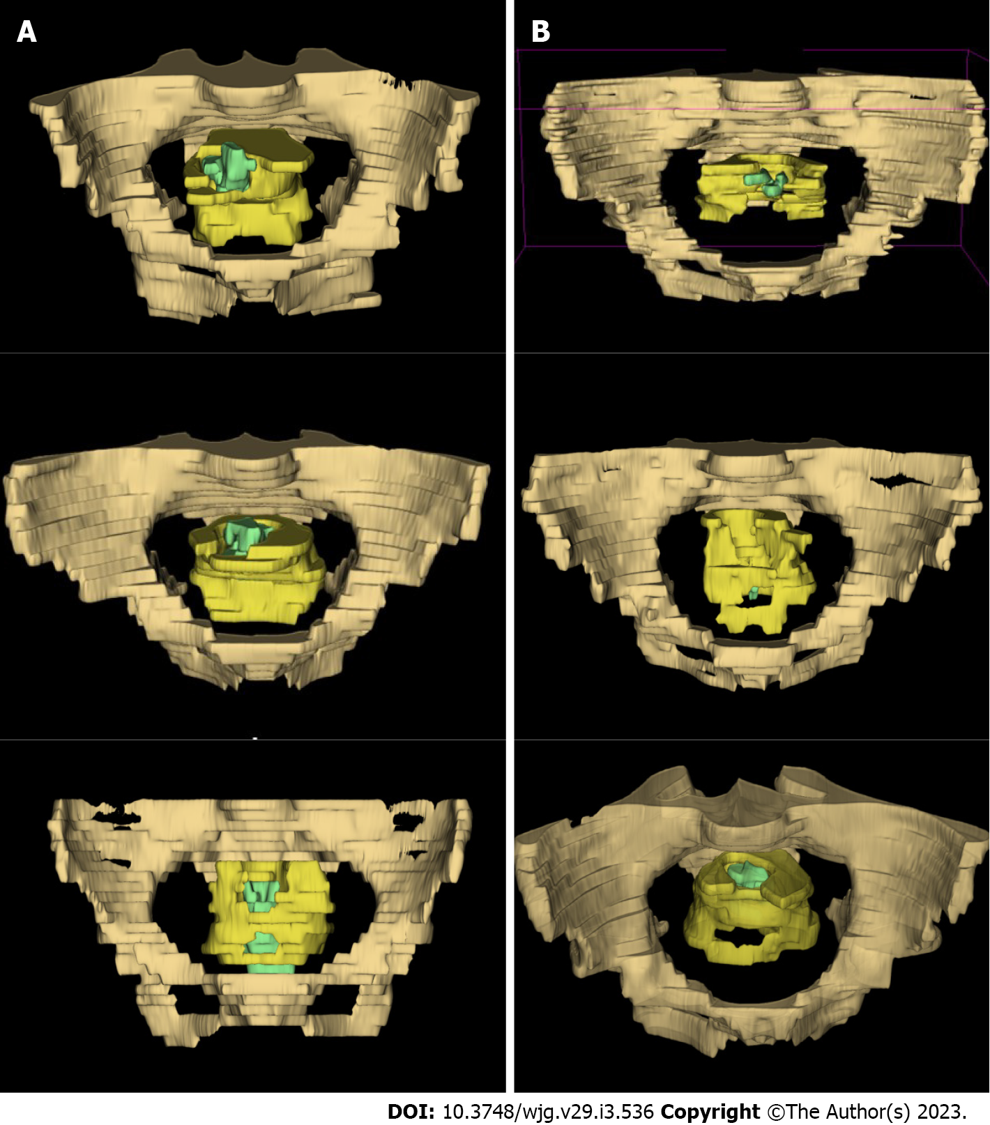
Figure 1 Examples of three-dimensional model of the target regions.
A: Models from patients with the use of ≥ 3 linear stapler cartridges; B: Models from patients with the use of ≤ 2 cartridges. The regions of pelvis, mesorectum, and tumor body were represented by drab, yellow, and green, respectively.
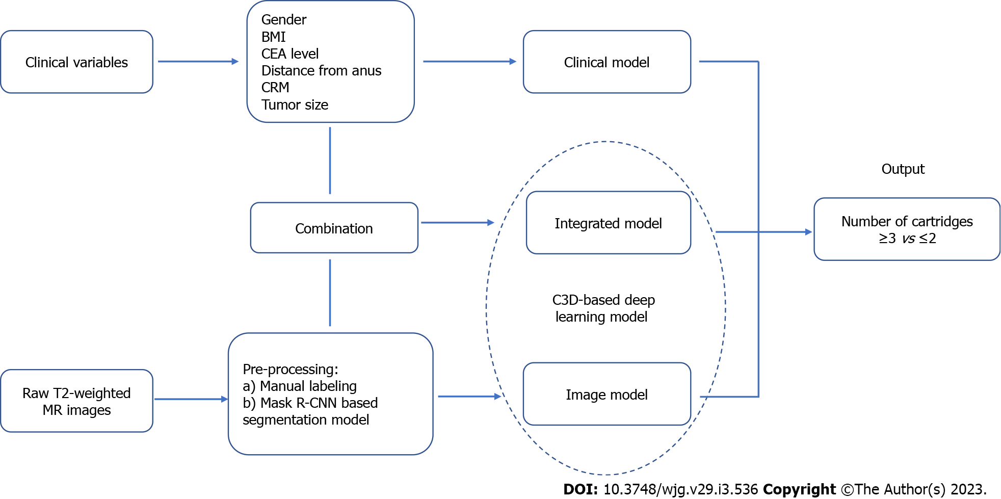
Figure 2 Flow chart of the design of pre-warning models.
BMI: Body mass index; CEA: Carcinoembryonic antigen; CRM: Circumferential resection margin; MR: Magnetic resonance; Mask R-CNN: Mask region-based convolutional neural network.
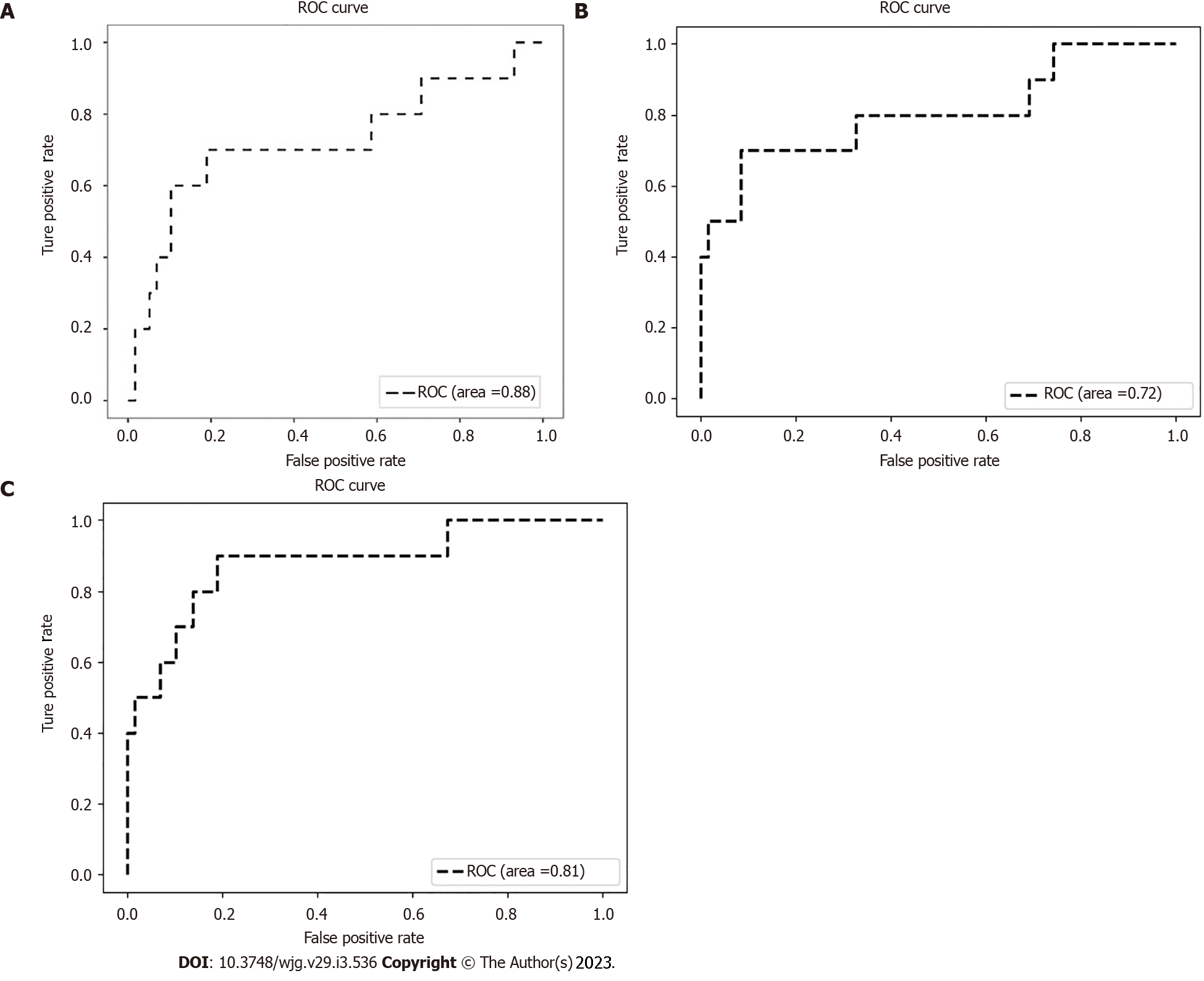
Figure 3 Receiver operating characteristic curves of the pre-warning models.
A: Clinical model; B: Image model; C: Integrated model.
- Citation: Cai ZH, Zhang Q, Fu ZW, Fingerhut A, Tan JW, Zang L, Dong F, Li SC, Wang SL, Ma JJ. Magnetic resonance imaging-based deep learning model to predict multiple firings in double-stapled colorectal anastomosis. World J Gastroenterol 2023; 29(3): 536-548
- URL: https://www.wjgnet.com/1007-9327/full/v29/i3/536.htm
- DOI: https://dx.doi.org/10.3748/wjg.v29.i3.536


