Copyright
©The Author(s) 2023.
World J Gastroenterol. May 7, 2023; 29(17): 2600-2615
Published online May 7, 2023. doi: 10.3748/wjg.v29.i17.2600
Published online May 7, 2023. doi: 10.3748/wjg.v29.i17.2600
Figure 1 Basic maneuvers of polypectomy.
A: Positioning the polyp at 5-6 o’clock on the monitor before polypectomy; B: The polyp’s base lying on gravity, this leads to poor visualization of the resection area; therefore, the patient’s position should be altered; C: The polyp’s base lying opposite to gravity after changing the patient’s position. This position change allows polyp stretching and avoids blood pooling on the resected site.
Figure 2 Endoloop-assisted polypectomy.
A: A pedunculated polyp with a thick stalk; B: Endoloop tightening around the stalk before snaring, the polyp’s color turned deep purple due to the ischemia; C: No bleeding evidence after the resection.
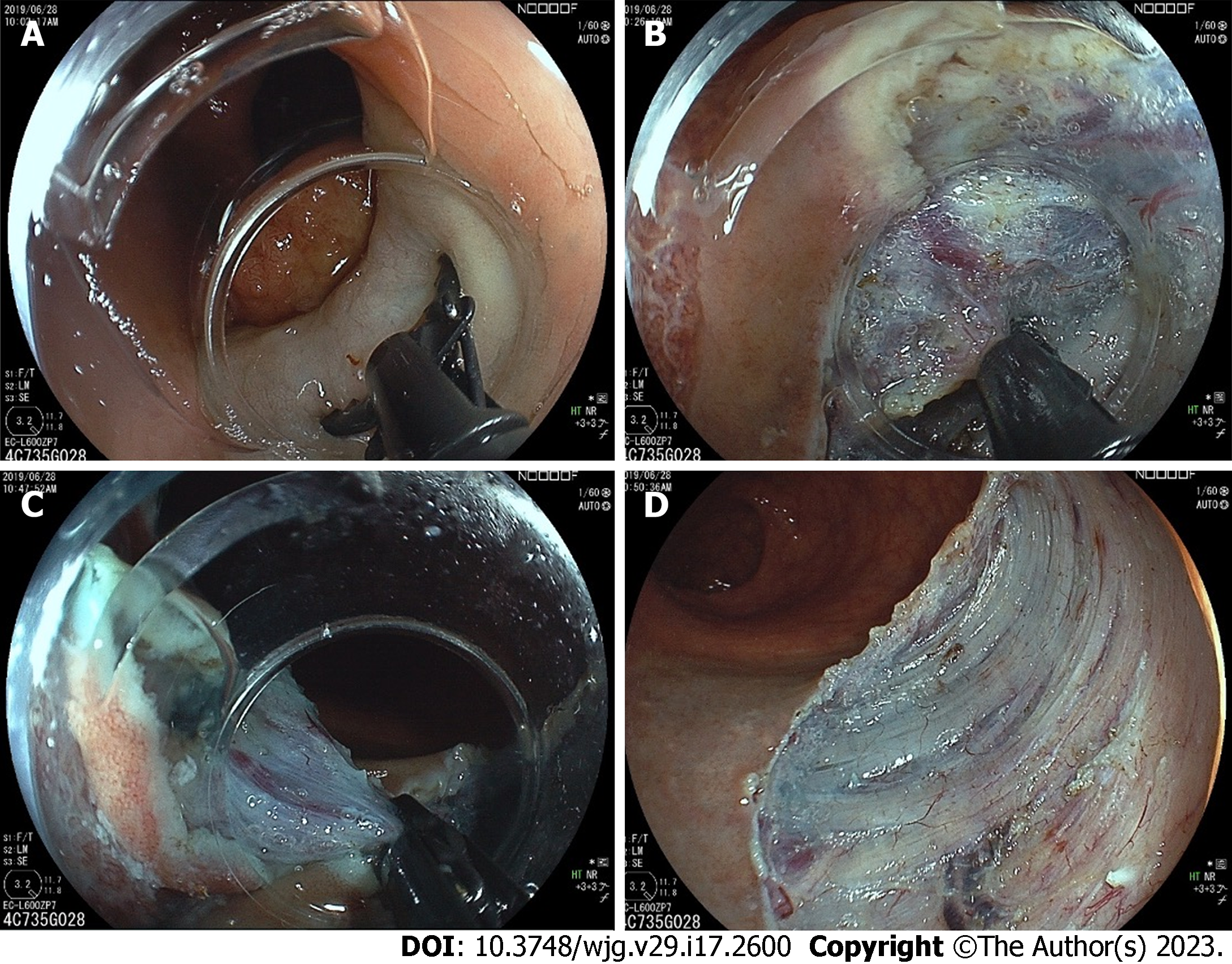
Figure 3 Endoscopic submucosal dissection with a scissor-type knife for a large subpedunculated polyp.
A: After submucosal injection, a scissor-type knife could be used to perform mucosal incision; B: The tissue and vessels could be grasped between two blades and precoagulated before cutting; C: Final cut of the procedure; D: Mucosal defect after completed endoscopic submucosal dissection.
Figure 4 Prophylactic clip application at the stalk before polypectomy.
A: A pedunculated polyp with a thick stalk and large head. There were failed attempts placing the endoloop due to the large polyp head; B: Clip application before snaring; C: No bleeding evidence after the resection.
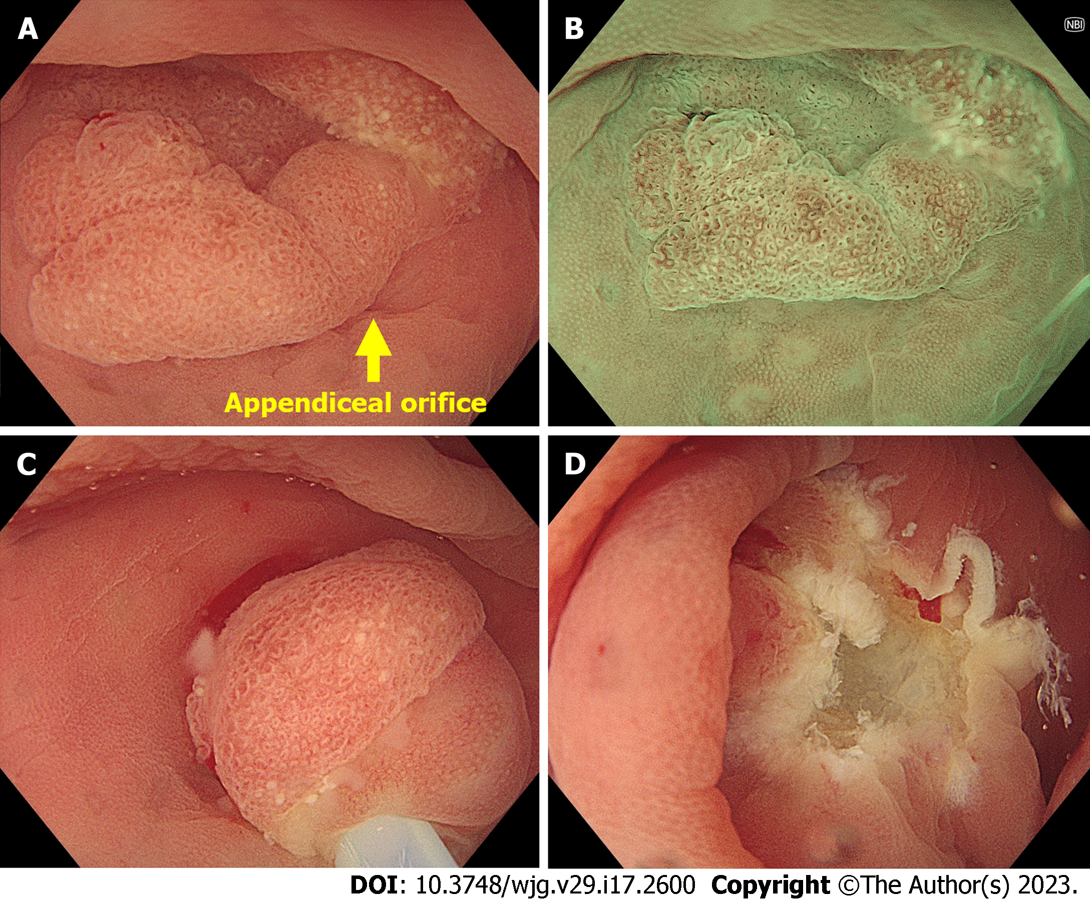
Figure 5 Underwater endoscopic mucosal resection for a polyp involving appendiceal orifice.
A: Endoscopic image showing a 15-mm sessile polyp involving the appendiceal orifice; B: Magnifying narrow-band image showing a type 1 polyp of the Japan Narrow-band Imaging Expert Team classification with open pit pattern. The most likely diagnosis was sessile serrated lesion; C: Underwater snaring without submucosal injection; D: Mucosal defect after completed underwater endoscopic mucosal resection.
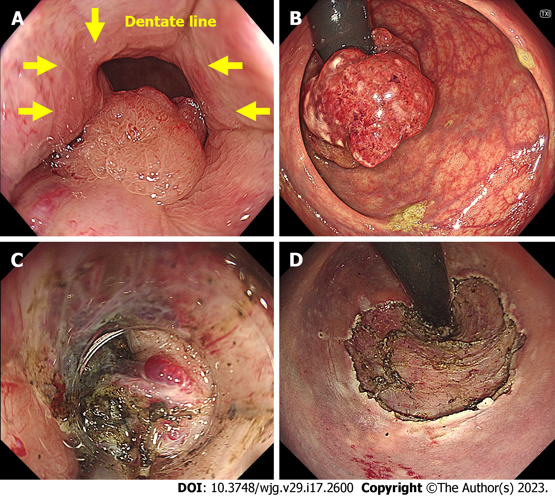
Figure 6 Endoscopic submucosal dissection for a polyp involving the anorectal junction.
A: Endoscopic image showing a 6-cm laterally spreading tumor involving the dentate line; B: Retroflexed view of the same polyp; C: Hemorrhoidal plexus at the anus makes the endoscopic submucosal dissection (ESD) challenging with the bleeding risk; D: Mucosal defect after completed ESD.
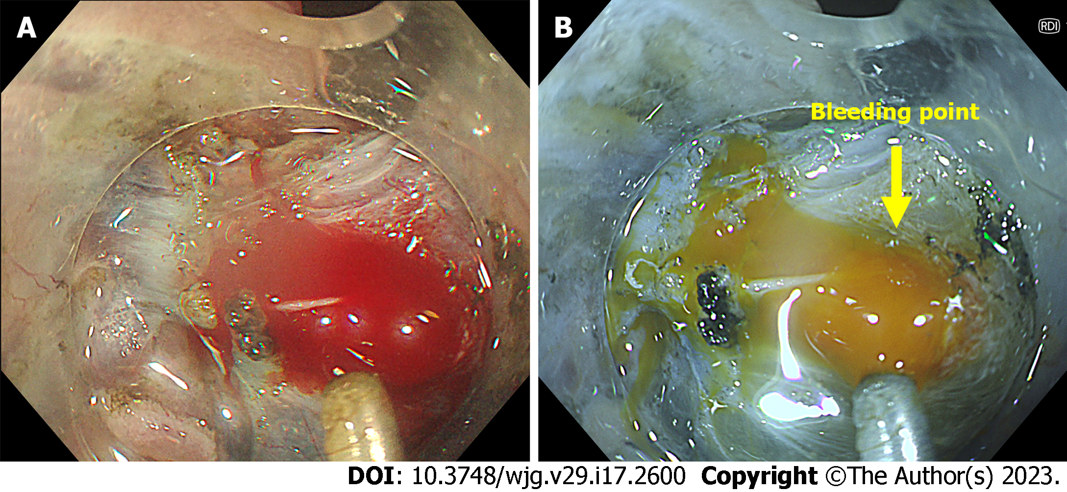
Figure 7 Bleeding during endoscopic submucosal dissection.
A: Endoscopic image in white light imaging showing the bleeding from the large vessels of the polyp; B: Identification of bleeding sources during polypectomy is easier in the Red Dichromatic Imaging (RDI) mode. RDI mode also reduces the endoscopists’ psychological stress by turning the blood color yellow.
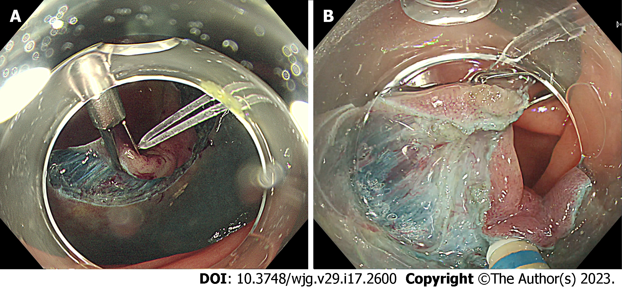
Figure 8 Traction device during endoscopic submucosal dissection.
A: Rubber band traction clip facilitating endoscopic submucosal dissection of a laterally spreading tumor with submucosal fibrosis; B: Traction force from the device helping during the submucosal dissection.
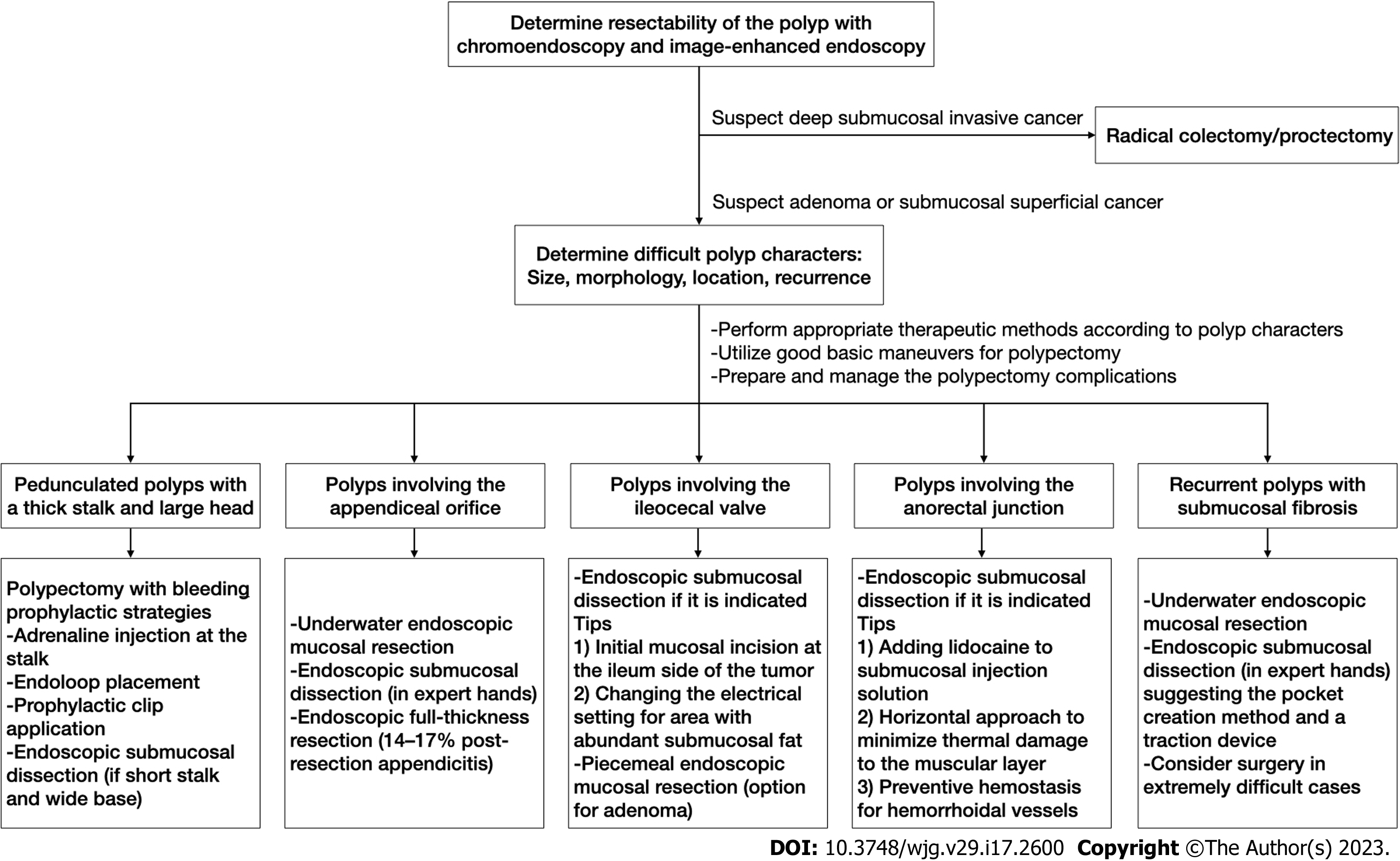
Figure 9
Stepwise approach for difficult colorectal polyps.
- Citation: Pattarajierapan S, Takamaru H, Khomvilai S. Difficult colorectal polypectomy: Technical tips and recent advances. World J Gastroenterol 2023; 29(17): 2600-2615
- URL: https://www.wjgnet.com/1007-9327/full/v29/i17/2600.htm
- DOI: https://dx.doi.org/10.3748/wjg.v29.i17.2600


