Copyright
©The Author(s) 2022.
World J Gastroenterol. Aug 7, 2022; 28(29): 3825-3837
Published online Aug 7, 2022. doi: 10.3748/wjg.v28.i29.3825
Published online Aug 7, 2022. doi: 10.3748/wjg.v28.i29.3825
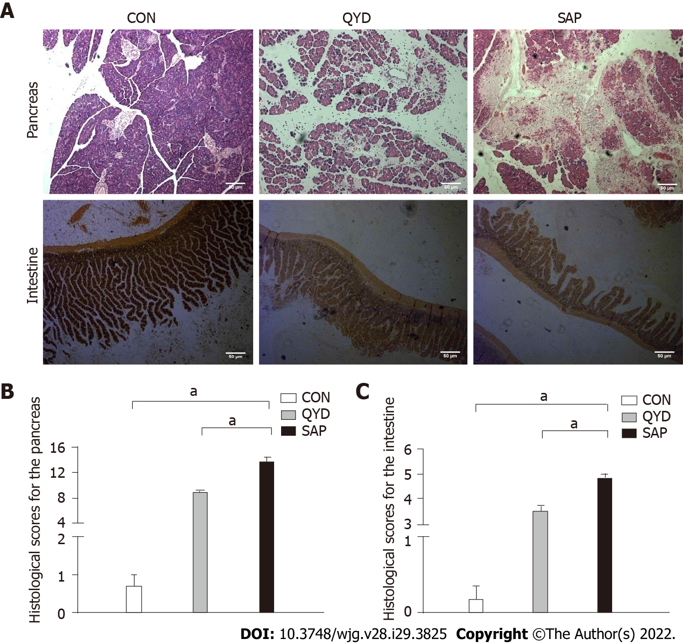
Figure 1 Histological changes in pancreatic and intestinal tissues.
A: Slight edema was detected [pancreas control (CON) group]; broad necrosis of acinar cells and interstitial edema were observed [pancreas severe acute pancreatitis (SAP) group]; only local necrosis, slight interstitial edema, and few inflammatory cell infiltrates were observed [pancreas Qingyi decoction (QYD) group]. Slight interstitial edema was detected (intestine CON group); irregular villi, mucosal erosion, and inflammatory infiltration were observed (intestine SAP group); edema and mild mucosal erosion were observed (intestine QYD group); B: Histological scores for the pancreas; C: Histological scores for the intestine. The histological scores for the pancreas and intestine were significantly ameliorated in the QYD treatment group compared with the SAP group. aP < 0.05, 100 ×. CON: Control; QYD: Qingyi decoction; SAP: Severe acute pancreatitis.
Figure 2 Serum levels in the different experimental groups.
A: Amylase; B: Interleukin-6; C: Tumor necrosis factor-α. aP < 0.05. CON: Control; QYD: Qingyi decoction; SAP: Severe acute pancreatitis; TNF: Tumor necrosis factor; IL: Interleukin.
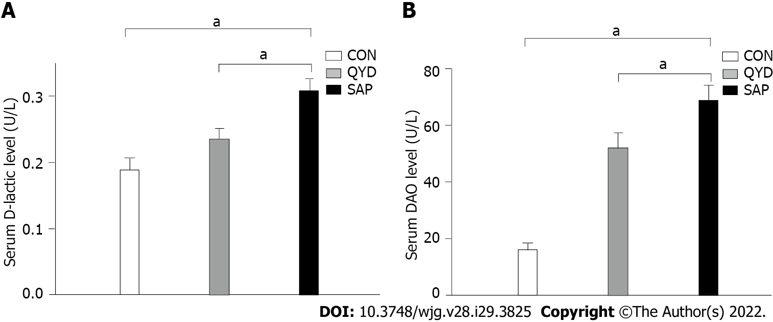
Figure 3 Effect of Qingyi decoction on serum.
A: D-lactic acid; B: Diamine oxidase. aP < 0.05. CON: Control; QYD: Qingyi decoction; SAP: Severe acute pancreatitis; DAO: Diamine oxidase.
Figure 4 Expression of calcineurin subunits in the intestine.
A: Calcineurin A (can) and calcineurin B (CnB) protein expression in the intestine; B: Qingyi decoction (QYD) treatment significantly inhibited the elevatcanCnA protein expression in the intestine observed in the severe acute pancreatitis (SAP) group; C: QYD treatment significantly inhibited the elevated CnB protein expression in the intestine observed in the SAP group. aP < 0.05. CON: Control; QYD: Qingyi decoction; SAP: Severe acute pancreatitis; CnA: Calcineurin A; CnB: Calcineurin B.
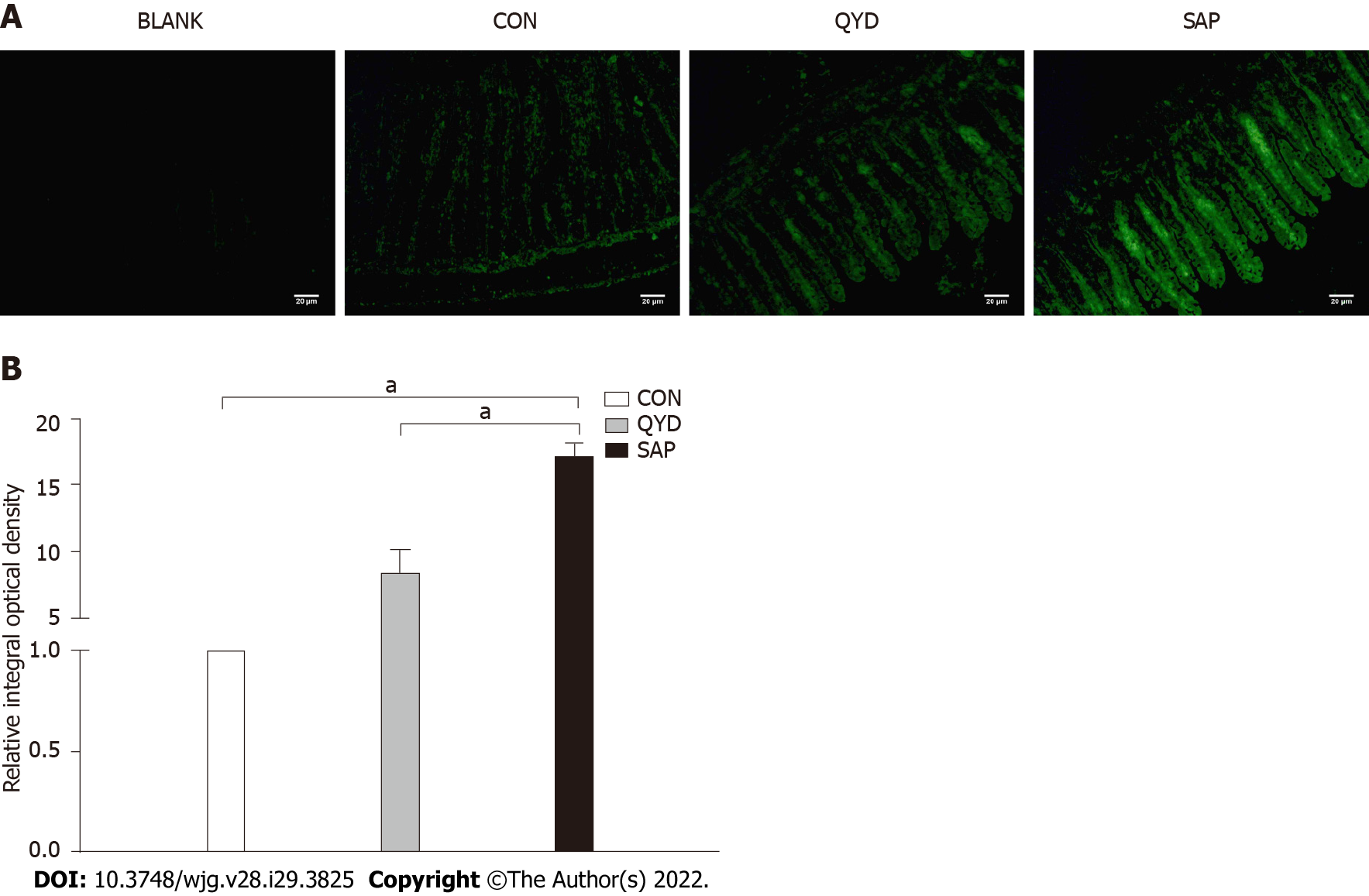
Figure 5 Immunofluorescence and semiquantitative analysis of nuclear factor of activated T-cells c3 expression.
A: Immunofluorescence of nuclear factor of activated T-cells c3 (NFATc3) expression; B: Semiquantitative analysis of NFATc3 expression. NFATc3 was highly expressed in the severe acute pancreatitis group compared with the control group but significantly downregulated under Qingyi decoction treatment. aP < 0.05. BLANK: Control without primary antibody; CON: Control; QYD: Qingyi decoction; SAP: Severe acute pancreatitis.
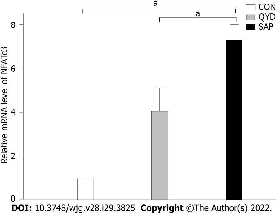
Figure 6 The level of nuclear factor of activated T-cells c3 mRNA transcription in intestinal tissue.
aP < 0.05. CON: Control; QYD: Qingyi decoction; SAP: Severe acute pancreatitis; NFATc3: Nuclear factor of activated T-cells c3.
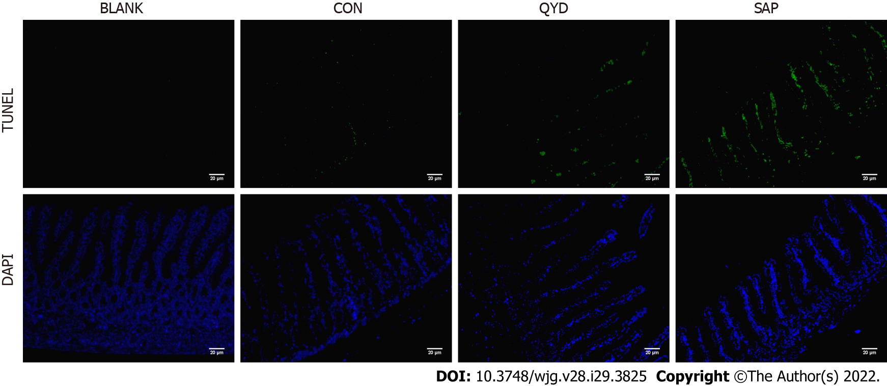
Figure 7 Transferase-mediated dUTP nick-end labeling assay to assess intestinal epithelial cell apoptosis.
Compared with that in the severe acute pancreatitis group, the level of intestinal epithelial cell apoptosis in the Qingyi decoction groups was significantly lower. 200 ×. BLANK: Control without primary antibody; CON: Control; QYD: Qingyi decoction; SAP: Severe acute pancreatitis. TUNEL: Transferase-mediated dUTP nick-end labeling; DAPI: 4’,6-diamidino-2-phenylindole.
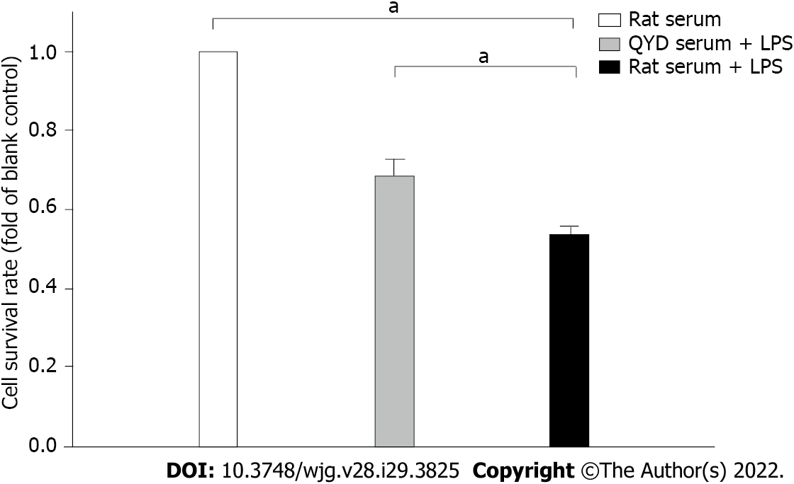
Figure 8 Effect of Qingyi decoction serum on lipopolysaccharide-induced cell viability.
aP < 0.05. QYD: Qingyi decoction; LPS: Lipopolysaccharide.
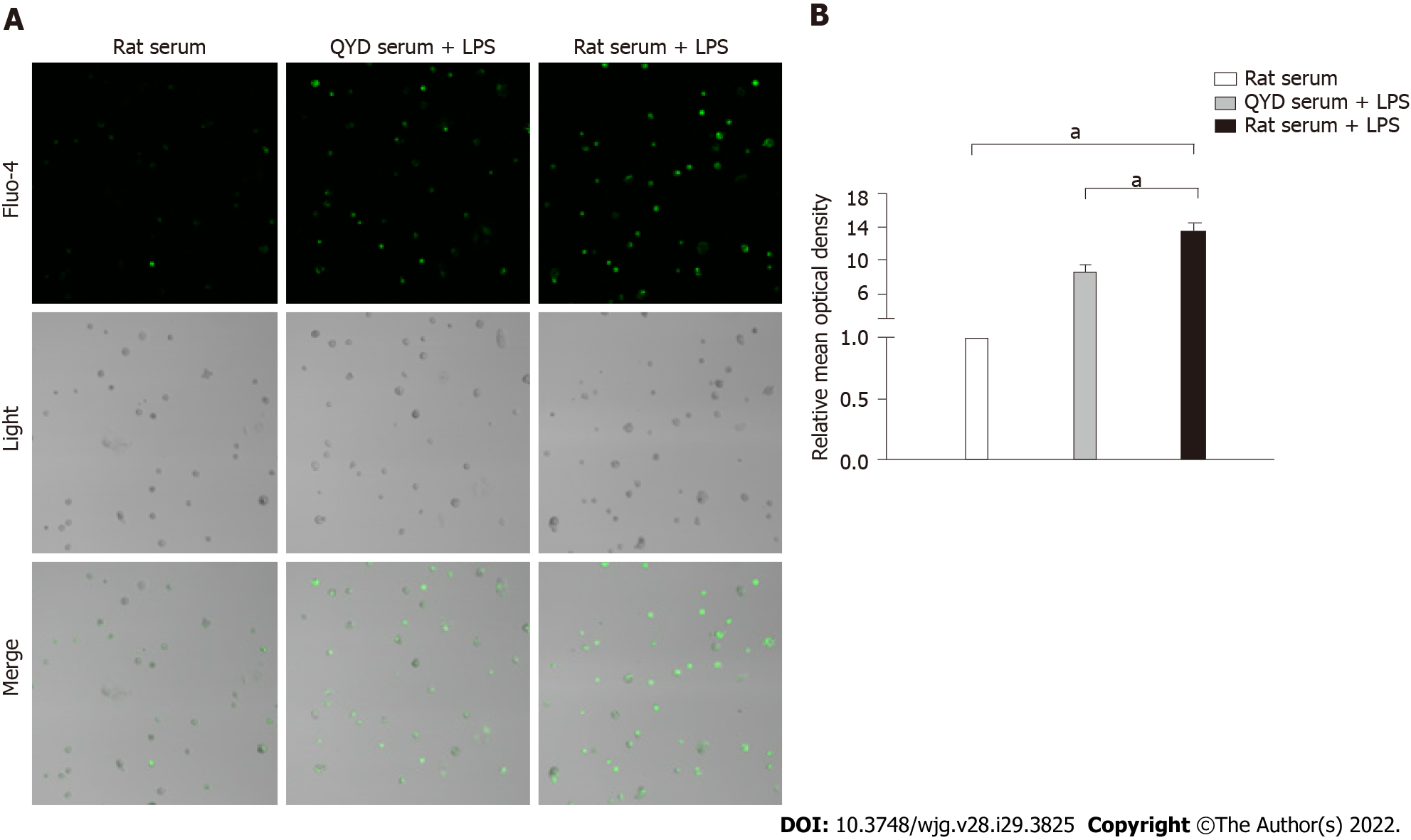
Figure 9 Fluorescence images of [Ca2+]i changes and the relative mean optical density.
A: Fluorescence images of Ca2+ changes measured using a confocal microscope; B: The relative mean optical density. Treatment with Qingyi decoction serum significantly attenuated the increase in Ca2+ levels observed in response to lipopolysaccharide-induced injury. aP < 0.05. QYD: Qingyi decoction; LPS: Lipopolysaccharide.
- Citation: Wang GY, Shang D, Zhang GX, Song HY, Jiang N, Liu HH, Chen HL. Qingyi decoction attenuates intestinal epithelial cell injury via the calcineurin/nuclear factor of activated T-cells pathway. World J Gastroenterol 2022; 28(29): 3825-3837
- URL: https://www.wjgnet.com/1007-9327/full/v28/i29/3825.htm
- DOI: https://dx.doi.org/10.3748/wjg.v28.i29.3825


