Copyright
©The Author(s) 2022.
World J Gastroenterol. Aug 7, 2022; 28(29): 3767-3779
Published online Aug 7, 2022. doi: 10.3748/wjg.v28.i29.3767
Published online Aug 7, 2022. doi: 10.3748/wjg.v28.i29.3767
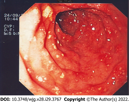
Figure 1 Endoscopic pictures of second portion of the duodenum.
The mucosa appears to be erythematous, hyperemic, and covered with diffuse erosions.
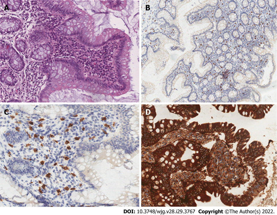
Figure 2 Spectrum of histologic findings in patients with intestinal involvement of systemic mastocytosis.
A: A mild initial finding with mast cells arranged in sheets and micro aggregates (hematoxylin-eosin, × 10). A heavy eosinophil infiltrate frequently dominates the picture. Crypt architectural distortion, without any other evidence of inflammatory bowel disease. Scattered lymphocytes and plasma cells often accompanied the mast cell and eosinophil infiltrates; B-C: The mast cells show expression of c-kit (B; CD117, × 2) and tryptase (C; × 10); D: CD25 positive cells in a clearcut and spread duodenal mastocytosis (CD25, × 10).
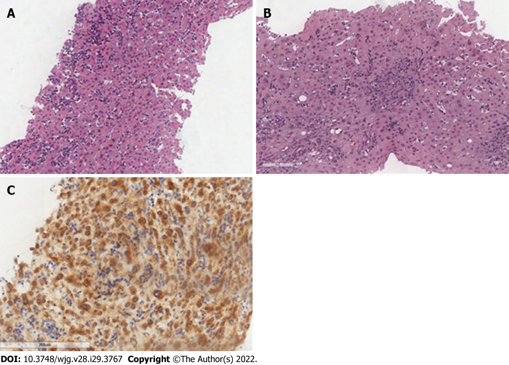
Figure 3 Frustules of hepatic parenchyma with substantially preserved structure, with trabeculae with 2-cell spinnerets with no significant steatosis, but focal balloon-like degeneration associated with biliary stasis phenomena.
A: Biliary spaces with preserved ducts, with ductular regeneration and bilio-hepatocyte metaplasia (hematoxylin-eosin, × 4); B: Some phenomena of hepatitis with aggression of the biliary epithelium are observed (hematoxylin-eosin, × 20). Portal spaces enlarged due to the presence of mixed inflammatory infiltrates, mainly consisting of T lymphocytes (CD3 +) with initial fibrotic expansion and formation of porto-portal bridges; C: In this context, scattered CD117 +, tryptase + cells (× 20) referable to mast cells.
- Citation: Elvevi A, Elli EM, Lucà M, Scaravaglio M, Pagni F, Ceola S, Ratti L, Invernizzi P, Massironi S. Clinical challenge for gastroenterologists–Gastrointestinal manifestations of systemic mastocytosis: A comprehensive review. World J Gastroenterol 2022; 28(29): 3767-3779
- URL: https://www.wjgnet.com/1007-9327/full/v28/i29/3767.htm
- DOI: https://dx.doi.org/10.3748/wjg.v28.i29.3767


