Copyright
©The Author(s) 2021.
World J Gastroenterol. Feb 28, 2021; 27(8): 692-707
Published online Feb 28, 2021. doi: 10.3748/wjg.v27.i8.692
Published online Feb 28, 2021. doi: 10.3748/wjg.v27.i8.692
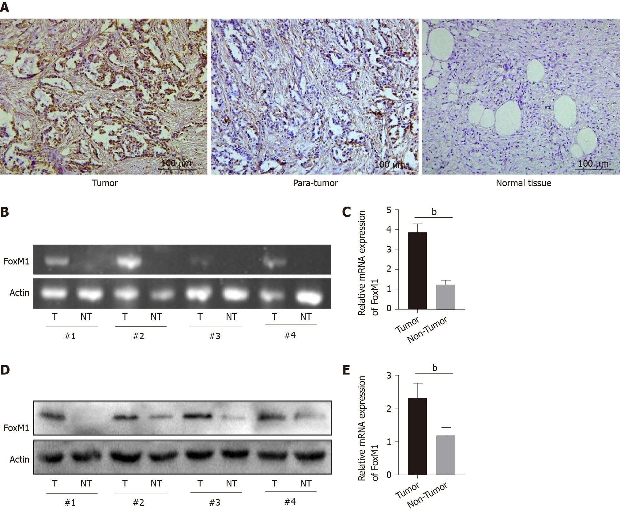
Figure 1 The expression of fork head box M1 in gallbladder cancer.
A: The expression of fork head box M1 in gallbladder cancer tissues (immunohistochemical staining, scale bars: 100 μm); B and C: Relative mRNA expression of fork head box M1; D and E: Relative protein expression of fork head box M1. aP < 0.05, bP < 0.01. FoxM1: Fork head box M1.
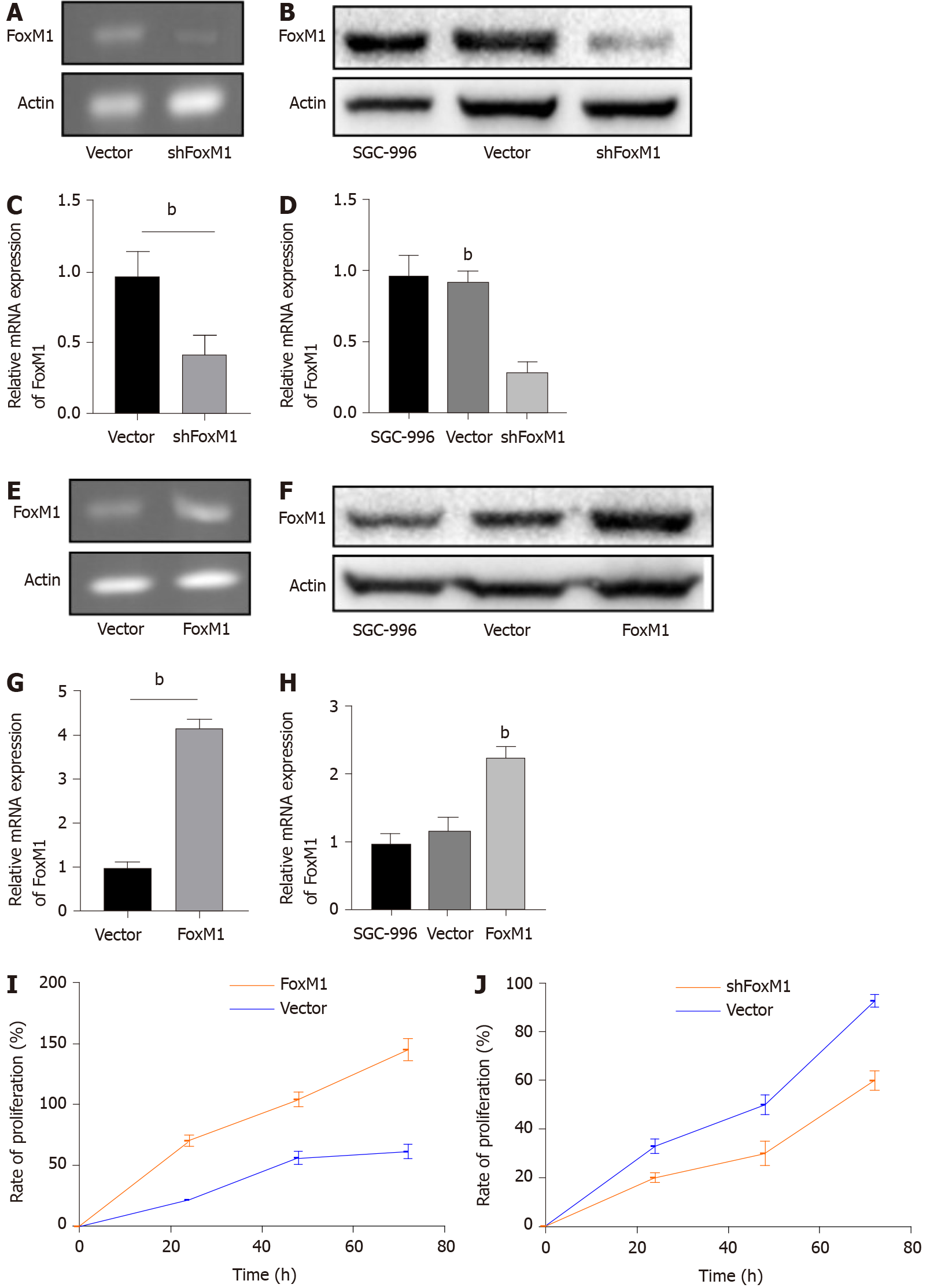
Figure 2 Fork head box M1 influenced proliferation of gallbladder cancer cells in vitro.
A-H: Relative mRNA and protein expression of fork head box M1; I: Rate of proliferation after overexpression of fork head box M1; J: Rate of proliferation after inhibition of fork head box M1. aP < 0.05, bP < 0.01. FoxM1: Fork head box M1.
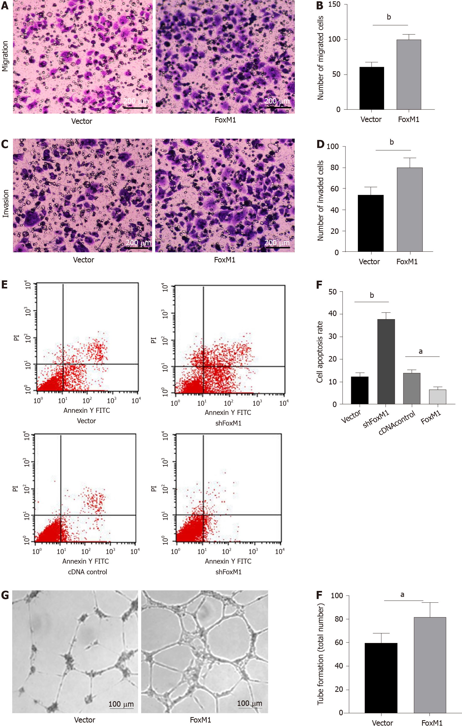
Figure 3 Fork head box M1 influenced migration and invasion of gallbladder cancer cells in vitro.
A-D: Lenti-fork head box M1-transfected cells had stronger migration and invasion abilities (cell invasion and migration assays, scale bars: 200 μm); E and F: Knockdown-fork head box M1 cells increased apoptotic levels (apoptosis assay); G and H: Upregulated fork head box M1 significantly enhanced vessel generation ability (endothelial cell tube formation assay, scale bars: 100 μm). aP < 0.05, bP < 0.01. FoxM1: Fork head box M1.
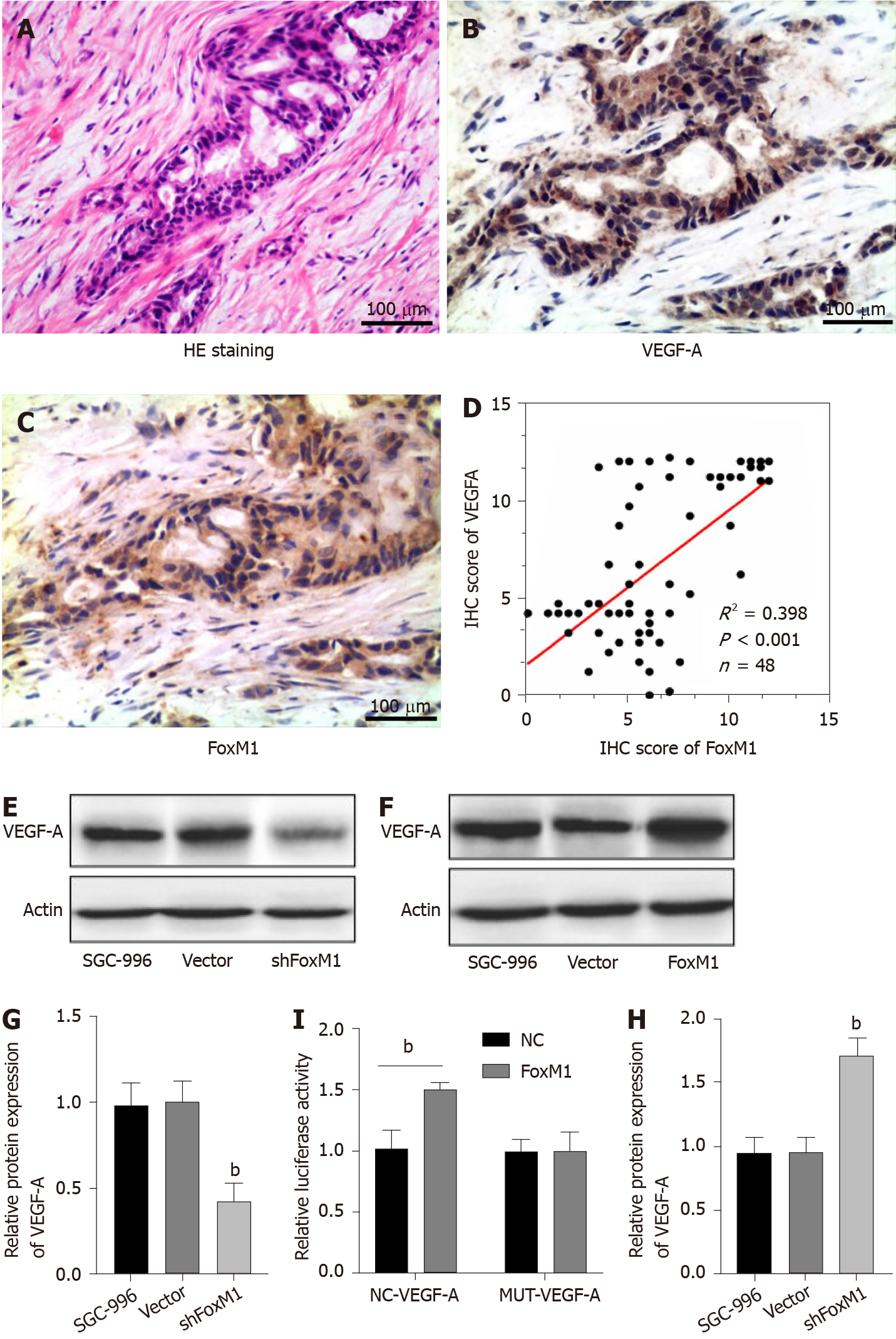
Figure 4 Fork head box M1 promoted gallbladder cancer cell proliferation and metastasis via vascular endothelial growth factor-A.
A-C: Hematoxylin-eosin staining, immunohistochemical staining of fork head box M1 and vascular endothelial growth factor-A (VEGF-A) in gallbladder cancer tissues (scale bars: 100 μm); D: Correlation between fork head box M1 and VEGF-A analyzed according to fluorescence score; E-H: Relative protein expression of VEGF-A; I: Luciferase assay of fork head box M1 and VEGF-A. aP < 0.05, bP < 0.01. FoxM1: Fork head box M1; VEGF-A: Vascular endothelial growth factor-A; NC: Normal control.
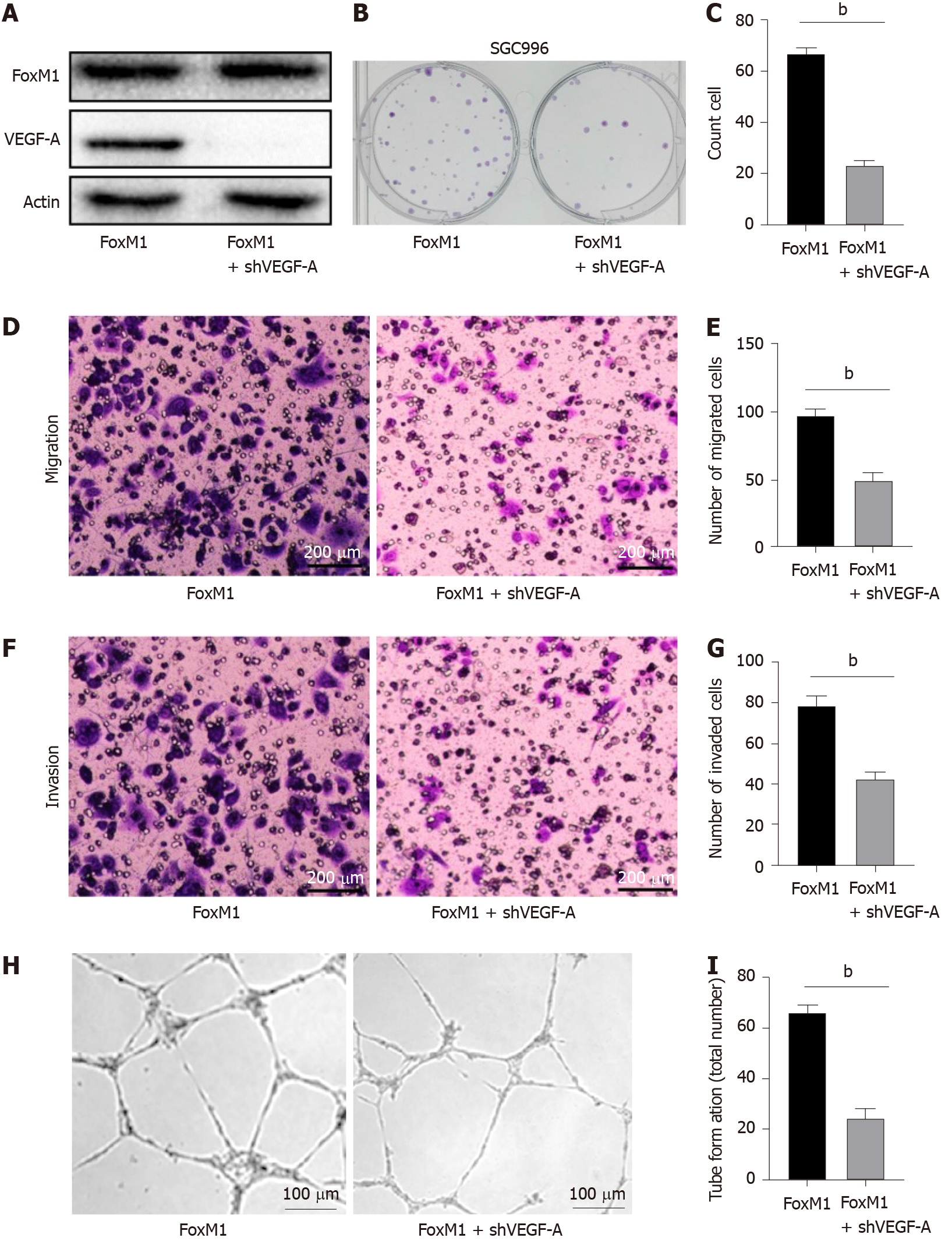
Figure 5 Knockdown vascular endothelial growth factor-A in fork head box M1 overexpressed cells could partly reverse the malignant phenotype of gallbladder cancer cells.
A: Relative protein expression of fork head box M1 and vascular endothelial growth factor-A; B and C: Clone formation of gallbladder cancer cells; D-G: Knockdown vascular endothelial growth factor-A in fork head box M1 overexpressed cells influenced migration and invasion abilities (cell invasion and migration assays, scale bars: 200 μm); H and I: Knockdown vascular endothelial growth factor-A in fork head box M1 overexpressed cells suppressed vessel generation ability (endothelial cell tube formation assay, scale bars: 100 μm). aP < 0.05, bP < 0.01. FoxM1: Fork head box M1; VEGF-A: Vascular endothelial growth factor-A.
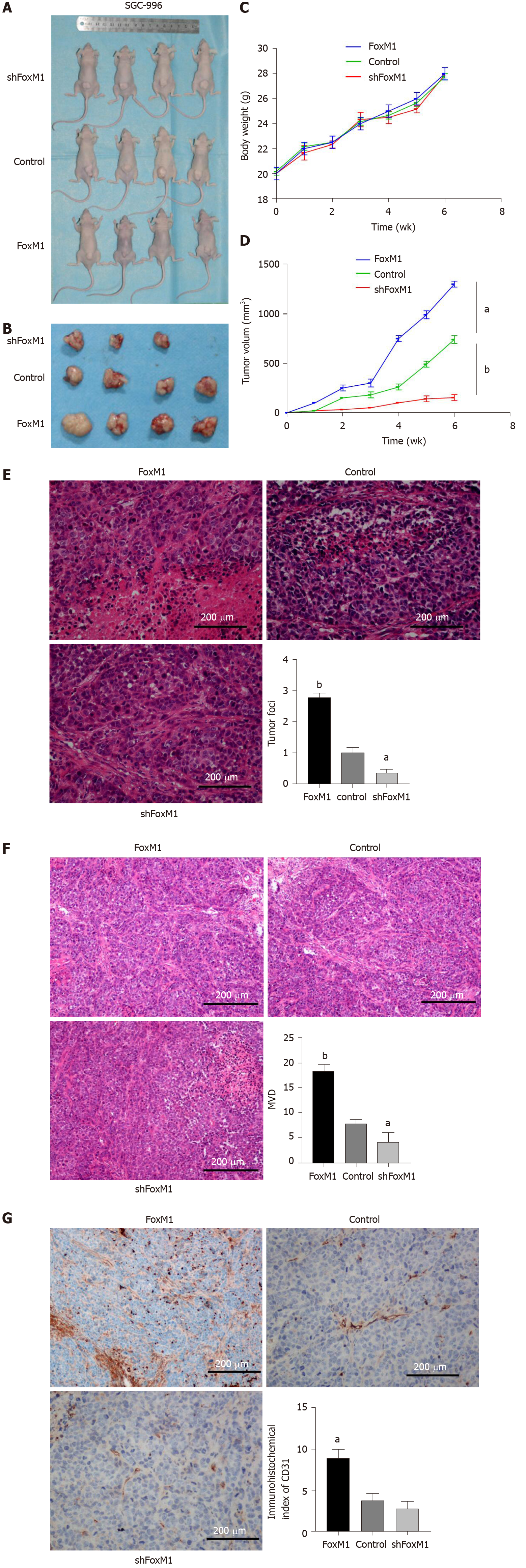
Figure 6 Fork head box M1 enhanced the angiogenesis of gallbladder cancer by regulating vascular endothelial growth factor-A in vivo.
A: Tumor-bearing mice [(1) Lenti-fork head box M1 (FoxM1)-shRNA, (2) cDNA-FoxM1, (3) control: xenotransplantation model]; B: Tumor from the three groups; C: Body weight of tumor-bearing mice; D: Tumor volume of tumor-bearing mice; E: Hematoxylin-eosin staining of tumor foci; F: Hematoxylin-eosin staining of microvascular density; G: Immunohistochemistry staining of CD31. Five fields per sample were counted; four samples in the control and FoxM1 groups and three samples in the shFoxM1 group were analyzed. Scale bars: 200 μm. aP < 0.05, bP < 0.01. FoxM1: Fork head box M1.
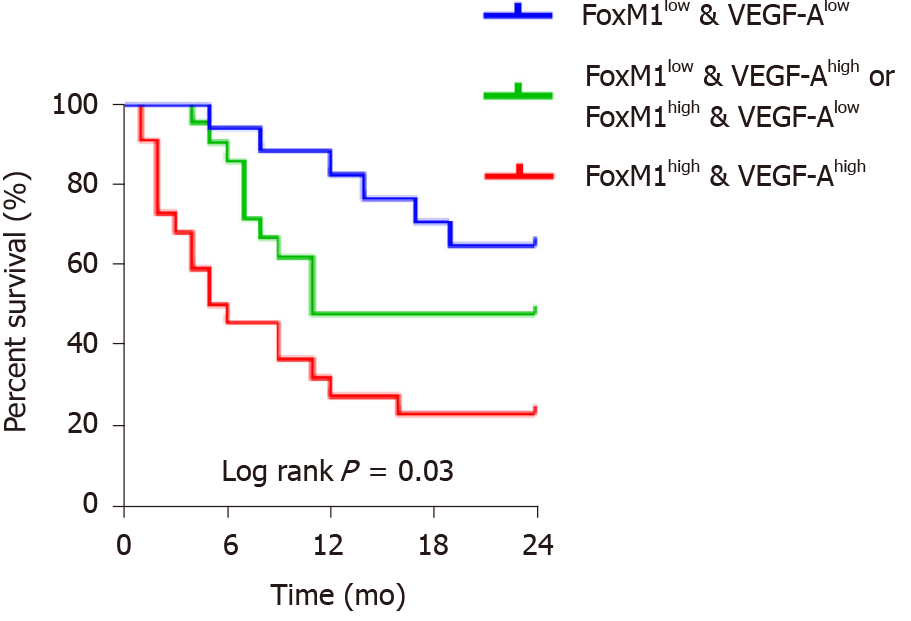
Figure 7 High expression of fork head box M1 and vascular endothelial growth factor-A were associated with poor prognosis of gallbladder cancer patients.
Kaplan-Meier analysis, n = Blue: 12, Green: 14, Red: 20. FoxM1: Fork head box M1; VEGF-A: Vascular endothelial growth factor-A.
- Citation: Wang RT, Miao RC, Zhang X, Yang GH, Mu YP, Zhang ZY, Qu K, Liu C. Fork head box M1 regulates vascular endothelial growth factor-A expression to promote the angiogenesis and tumor cell growth of gallbladder cancer. World J Gastroenterol 2021; 27(8): 692-707
- URL: https://www.wjgnet.com/1007-9327/full/v27/i8/692.htm
- DOI: https://dx.doi.org/10.3748/wjg.v27.i8.692


