Copyright
©The Author(s) 2021.
World J Gastroenterol. Feb 14, 2021; 27(6): 534-544
Published online Feb 14, 2021. doi: 10.3748/wjg.v27.i6.534
Published online Feb 14, 2021. doi: 10.3748/wjg.v27.i6.534
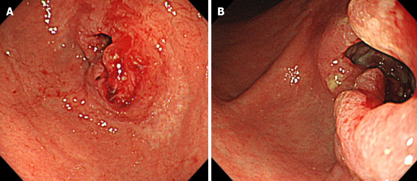
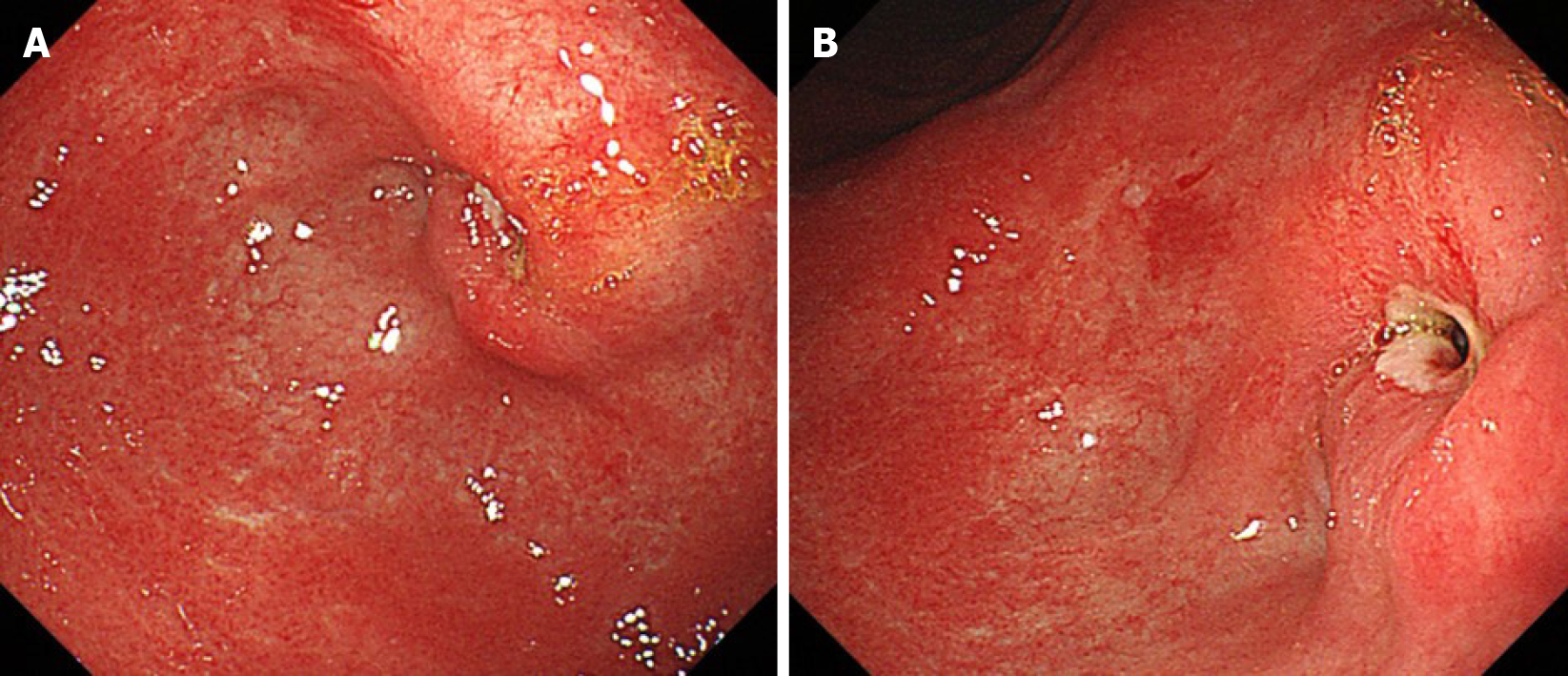
Figure 1 Initial endoscopic findings.
A and B: Type 3 primary tumor located on the lower part of the stomach.
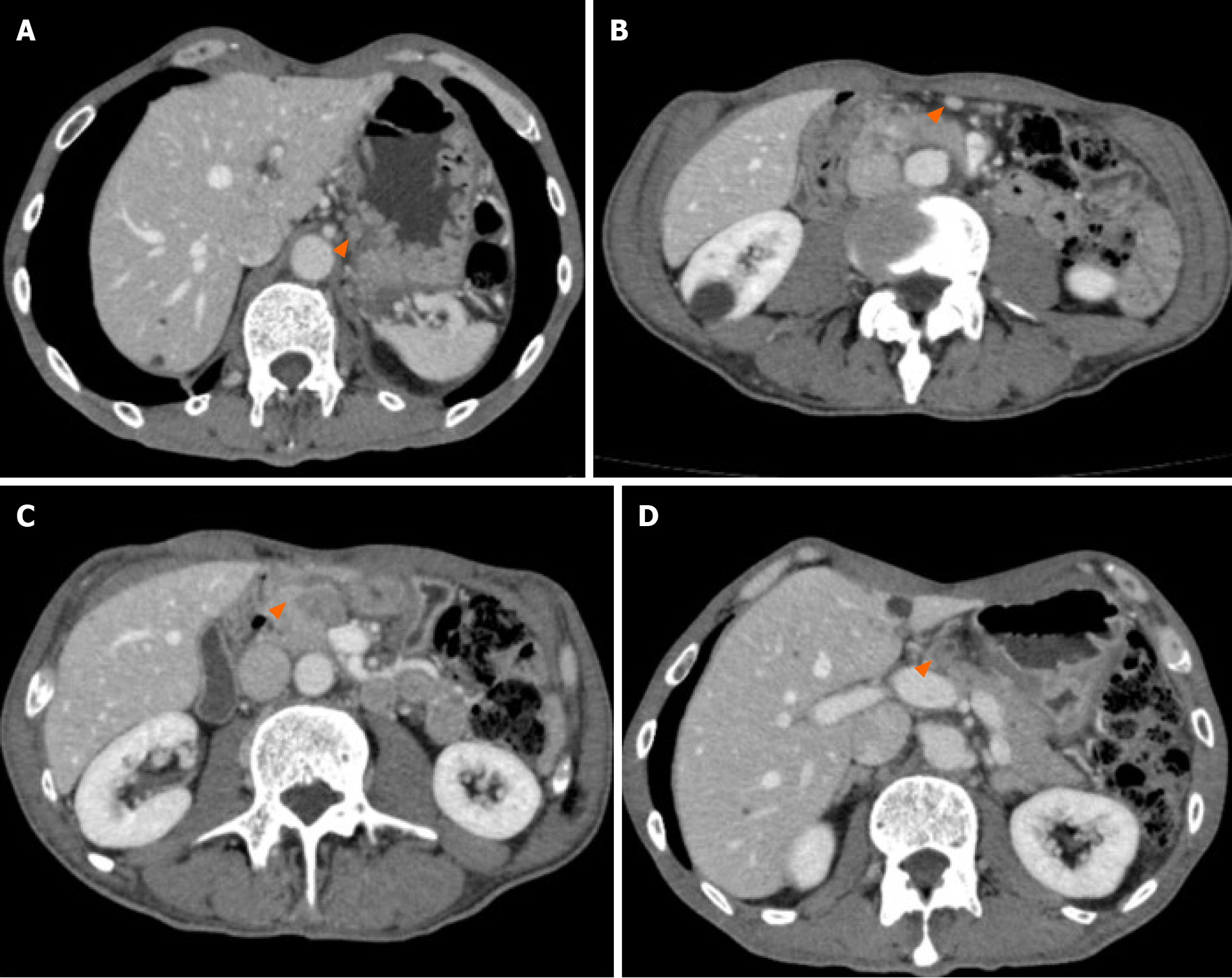
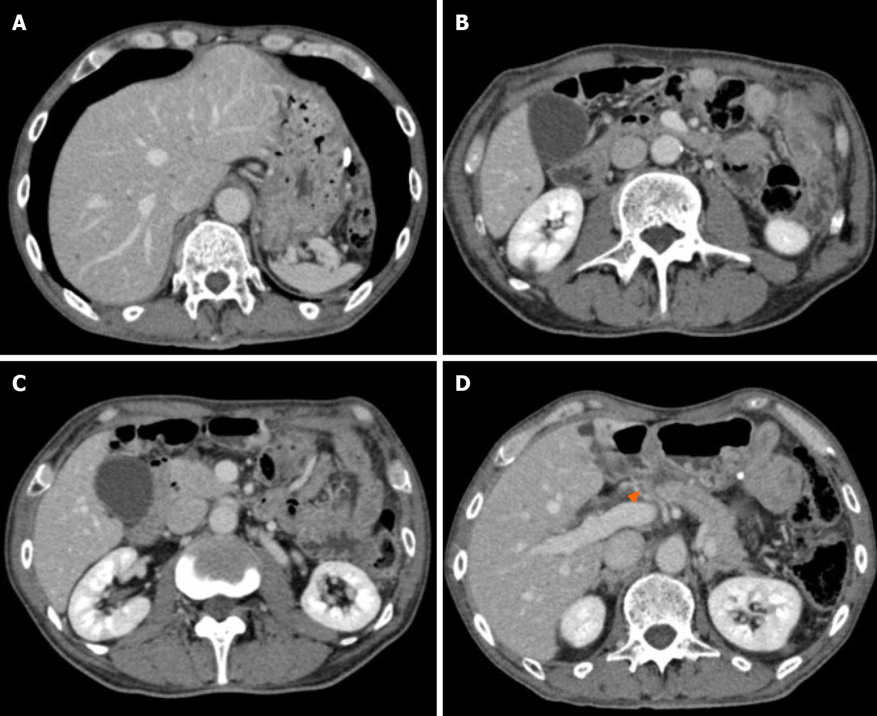
Figure 2 Initial computed tomography findings, computed tomography showed lymph node swelling.
A: Swelling of #3 lymph node (LN) (orange triangle; 11.8 mm × 8.5 mm); B: Swelling of #4d LN (orange triangle; 10.3 mm × 8.4 mm); C: Swelling of #6 LN, suspected to be invasion to the pancreatic head (orange angle; 21.6 mm × 14.7 mm); D: Swelling of #8a LN (orange triangle; 14.0 mm × 13.4 mm).
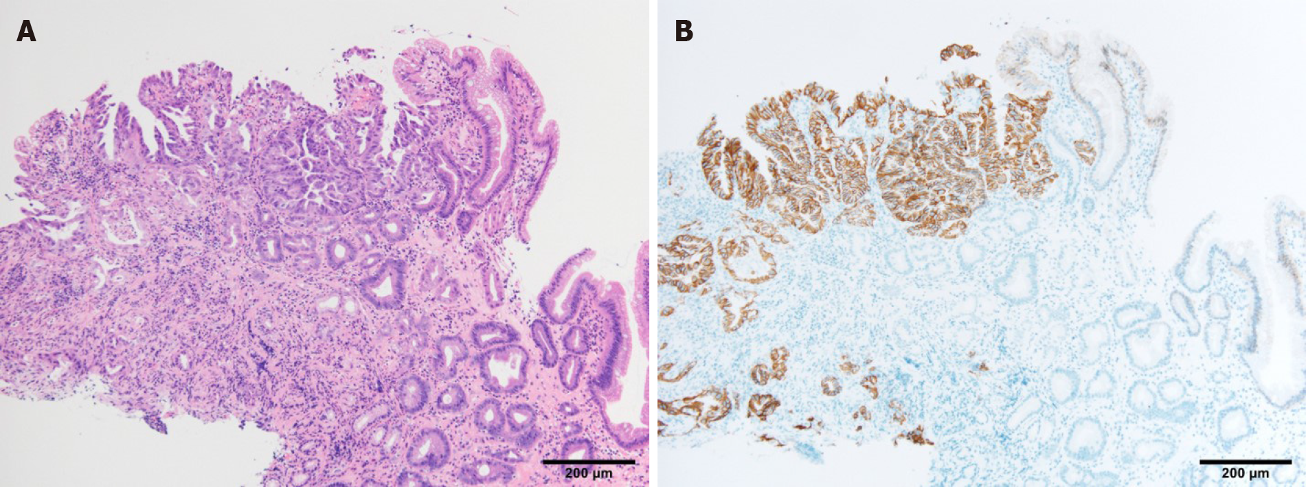
Figure 3 Pathological findings from endoscopic biopsy.
Pathological examination of the endoscopic biopsy revealed a papillary and well-differentiated adenocarcinoma. A: Hematoxylin and eosin staining results; × 10; B: Human epidermal growth factor receptor 2 positivity was detected by an immunohistochemical staining; × 10.
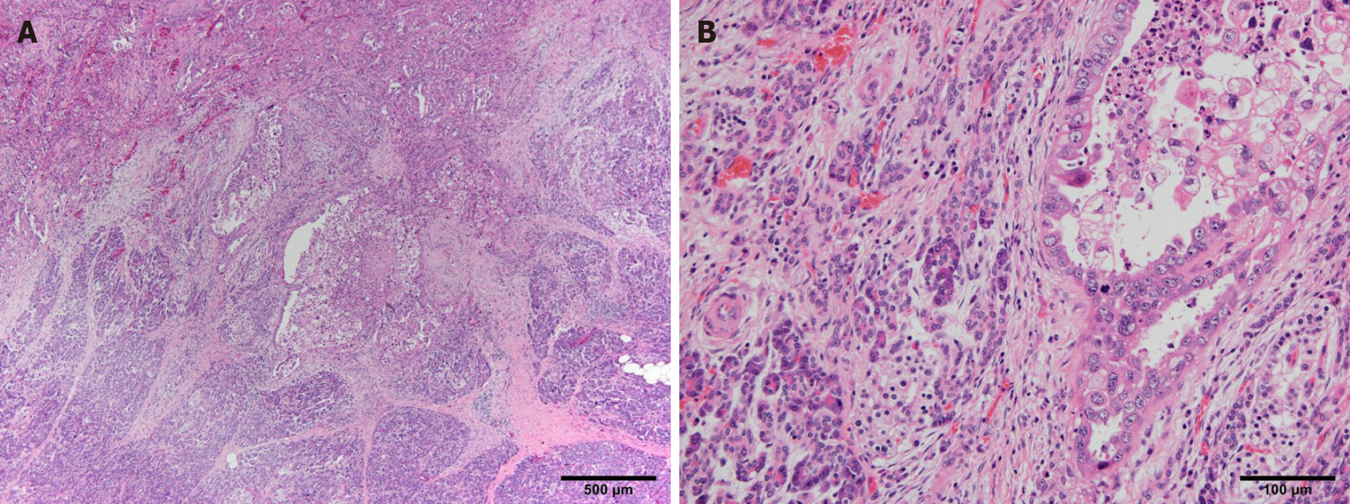
Figure 4 Pathological findings from resected specimens showing pancreatic infiltration of cancer cells.
A: Hematoxylin and eosin staining results; × 4; B: Hematoxylin and eosin staining results; × 20.
Figure 5 Endoscopic findings after neoadjuvant chemotherapy.
A and B Shrinkage of the primary tumor.
Figure 6 Computed tomography findings after neoadjuvant chemotherapy.
A: #3 lymph node (LN) became smaller and could not be detected; B: #4d LN became smaller and could not be detected; C: #6 LN became smaller and could not be detected; D: #8a LN became smaller (orange triangle; 14.0 mm × 13.4 mm to 9.2 mm × 7.0 mm).
- Citation: Yura M, Takano K, Adachi K, Hara A, Hayashi K, Tajima Y, Kaneko Y, Ikoma Y, Fujisaki H, Hirata A, Hongo K, Yo K, Yoneyama K, Dehari R, Koyanagi K, Nakagawa M. Pancreaticoduodenectomy after neoadjuvant chemotherapy for gastric cancer invading the pancreatic head: A case report. World J Gastroenterol 2021; 27(6): 534-544
- URL: https://www.wjgnet.com/1007-9327/full/v27/i6/534.htm
- DOI: https://dx.doi.org/10.3748/wjg.v27.i6.534


