Copyright
©The Author(s) 2021.
World J Gastroenterol. Feb 14, 2021; 27(6): 487-500
Published online Feb 14, 2021. doi: 10.3748/wjg.v27.i6.487
Published online Feb 14, 2021. doi: 10.3748/wjg.v27.i6.487
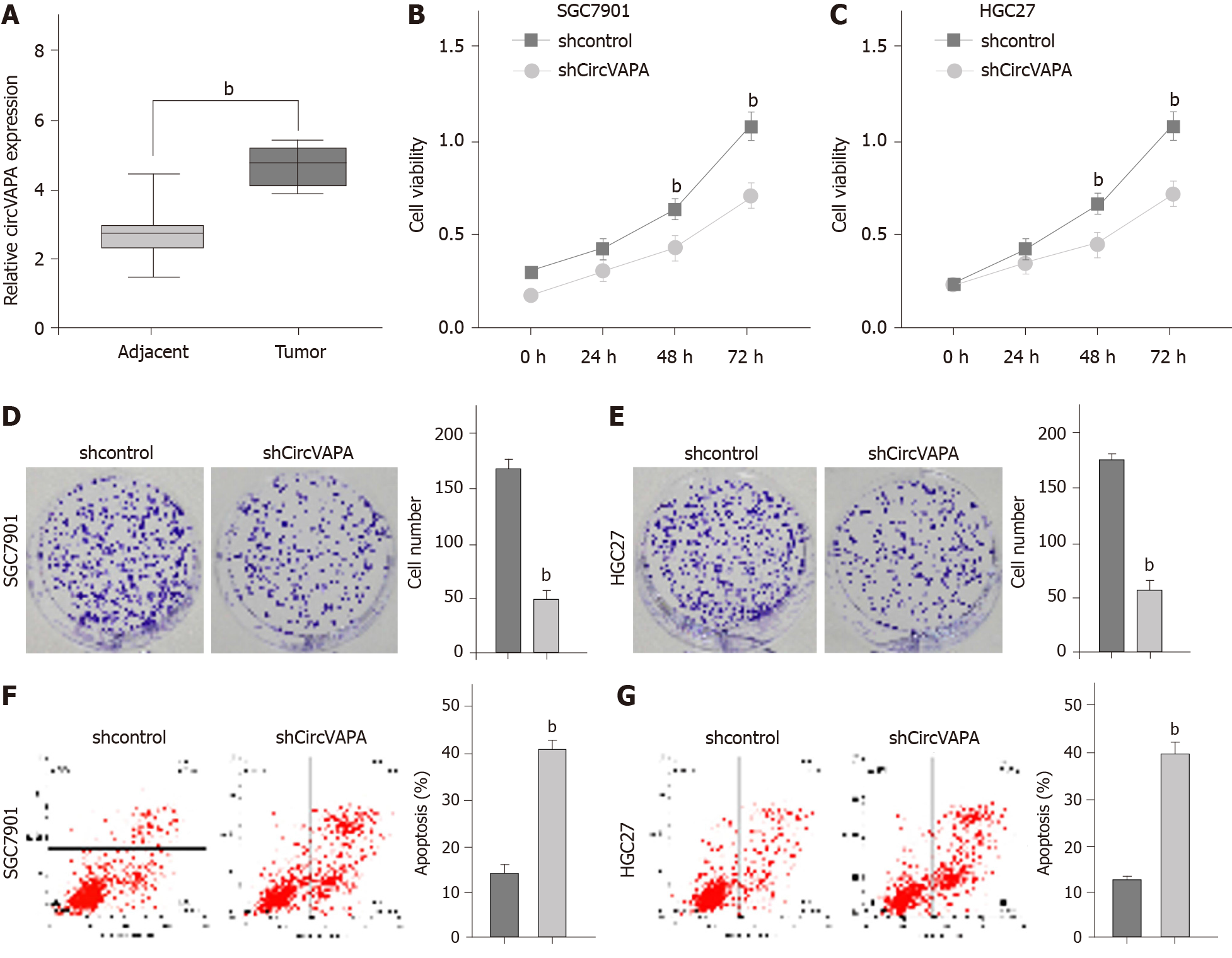
Figure 1 CircVAPA enhances proliferation and represses apoptosis of gastric cancer cells.
A: The expression of circVAPA was measured by qPCR in the gastric cancer tissues (n = 50) and adjacent normal tissues (n = 50); B-G: SGC7901 and HGC27 cells were treated with the circVAPA shRNA or control shRNA. Cell viability was tested by MTT assay (B and C), cell proliferation was measured by colony formation assay (D and E), and cell apoptosis was analyzed by flow cytometry (F and G). Data are presented as the mean ± SD. bP < 0.01.
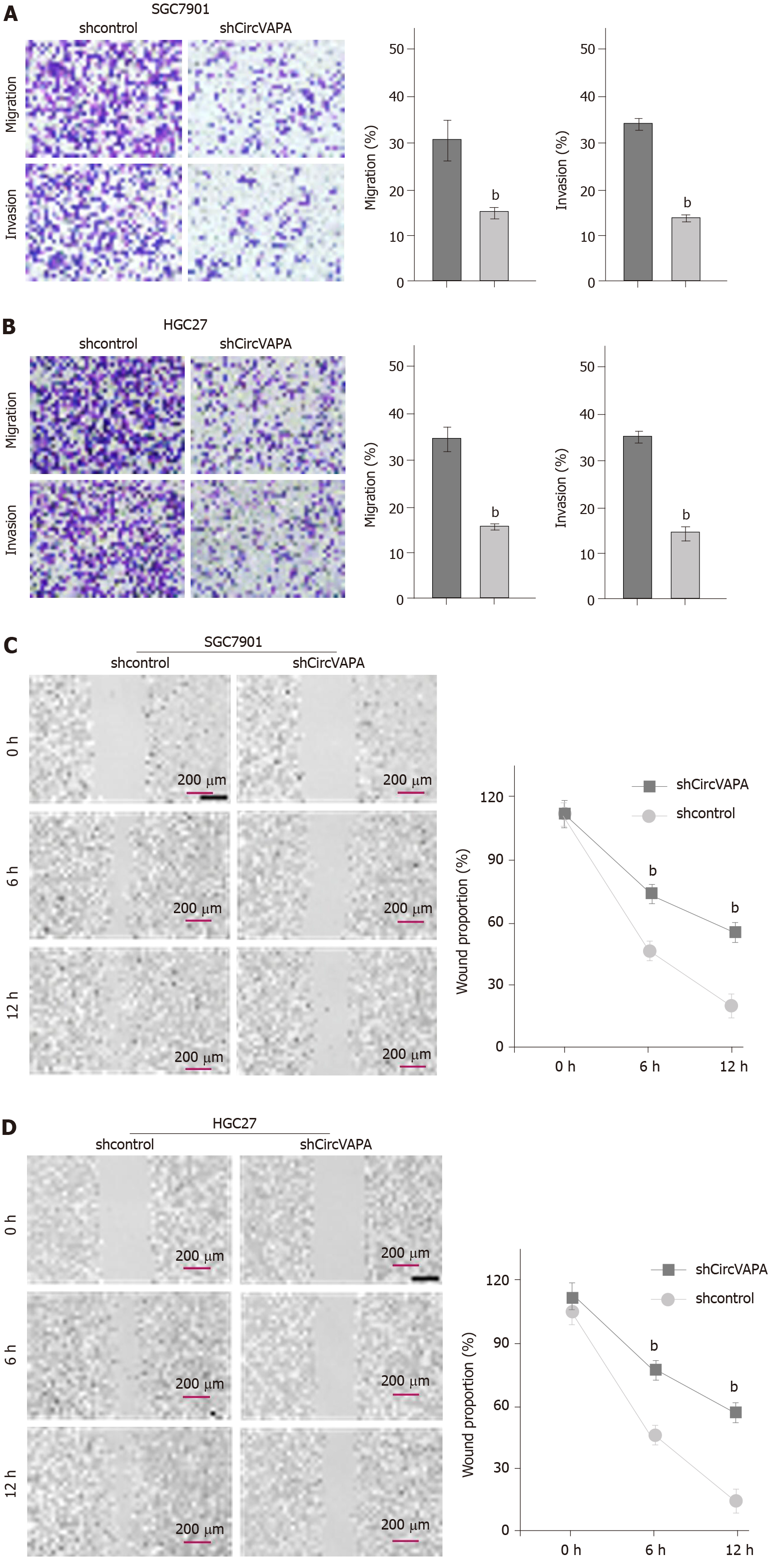
Figure 2 CircVAPA promotes invasion and migration of gastric cancer cells.
A-D: SGC7901 and HGC27 cells were treated with the circVAPA shRNA or control shRNA. Cell migration and invasion were tested by transwell assay (A and B), and migration and invasion were analyzed by wound healing assay (C and D). The wound healing proportion is shown. Data are presented as the mean ± SD. bP < 0.01.
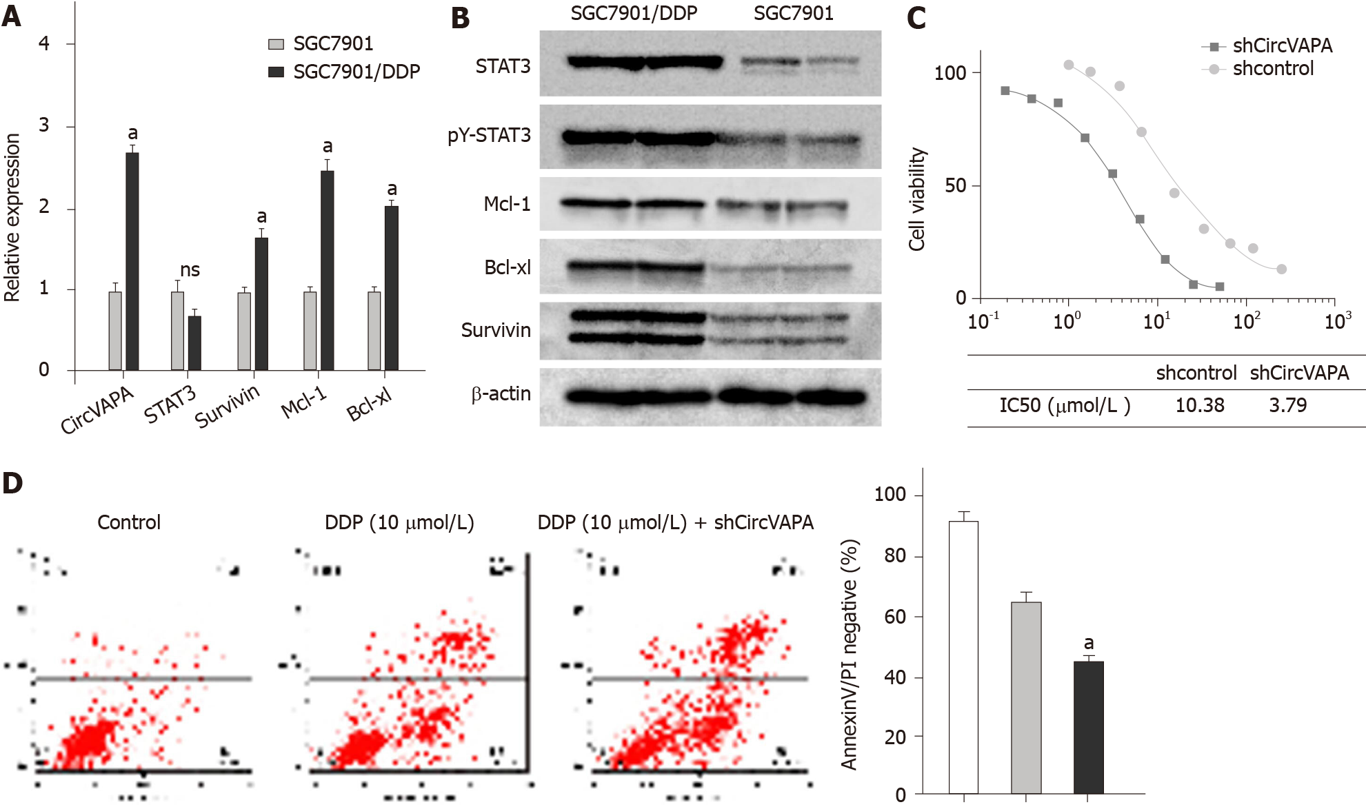
Figure 3 Elevation of CircVAPA enhances chemotherapy drug resistance of gastric cancer cells.
A: The expression of circVAPA, STAT3, Survivin, Mcl-1, and Bcl-xl was measured by qPCR in the SGC7901 and SGC7901/cisplatin (DDP) cells; B: STAT3 phosphorylation and levels of STAT3, Survivin, Mcl-1, and Bcl-xl were tested by Western blot analysis; C: SGC7901/DDP cells were initially treated with DDP at indicated doses and then with control shRNA or circVAPA shRNA. Cell viability was analyzed by MTT assay; D: SGC7901/DDP cells were treated with DDP or co-treated with DDP and circVAPA shRNA. Cell apoptosis was analyzed by flow cytometry. Data are presented as the mean ± SD. aP < 0.05.
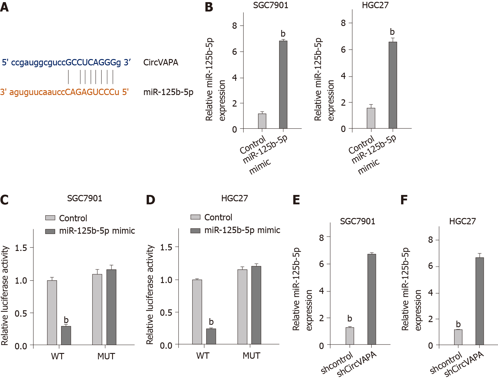
Figure 4 CircVAPA is able to sponge miR-125b-5p in gastric cancer cells.
A: The interaction of CircVAPA and miR-125b-5p was analyzed by bioinformatic analysis based on ENCORI (http://starbase.sysu.edu.cn/index.php); B-D: SGC7901 and HGC27 cells were treated with control mimic or miR-125b-5p mimic. The expression levels of miR-125b-5p were tested by qPCR in the cells (B), and luciferase activities of wild type CircVAPA and CircVAPA with the miR-125b-5p-binding site mutant were examined by luciferase reporter gene assay (C and D); E and F: SGC7901 and HGC27 cells were treated with the circVAPA shRNA or control shRNA. The expression of miR-125b-5p was analyzed by qPCR assay. Data are presented as the mean ± SD. bP < 0.01. MUT: Mutant; WT: Wild type.
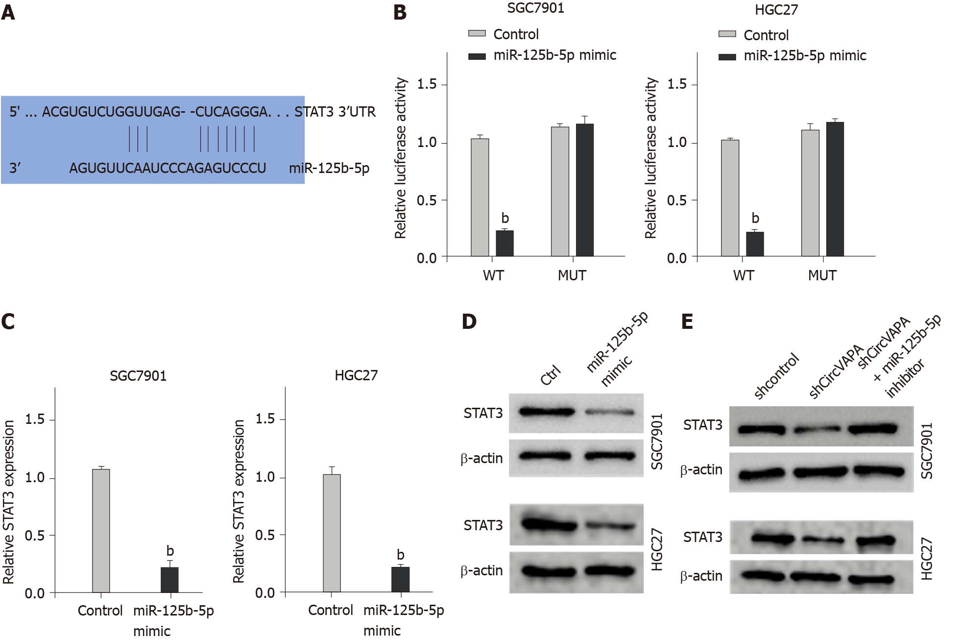
Figure 5 MiR-125b-5p targets STAT3 in gastric cancer cells.
A: The binding of miR-125b-5p and the 3’ untranslated region of STAT3 was identified by bioinformatic analysis based on Targetscan (http://www.targetscan.org/vert_72/); B-D: SGC7901 and HGC27 cells were treated with control mimic or miR-125b-5p mimic. The luciferase activities of wild type STAT3 and STAT3 with the miR-125b-5p-binding site mutant were examined by luciferase reporter gene assay (B), the mRNA expression of STAT3 was analyzed by qPCR assay (C), and the protein expression of STAT3 was determined by Western blot analysis (D); E: SGC7901 and HGC27 cells were treated with control shRNA or circVAPA shRNA, or co-treated with circVAPA shRNA and miR-125b-5p inhibitor. The protein expression of STAT3 was tested by Western blot analysis. Data are presented as the mean ± SD. bP < 0.01. MUT: Mutant; WT: Wild type.
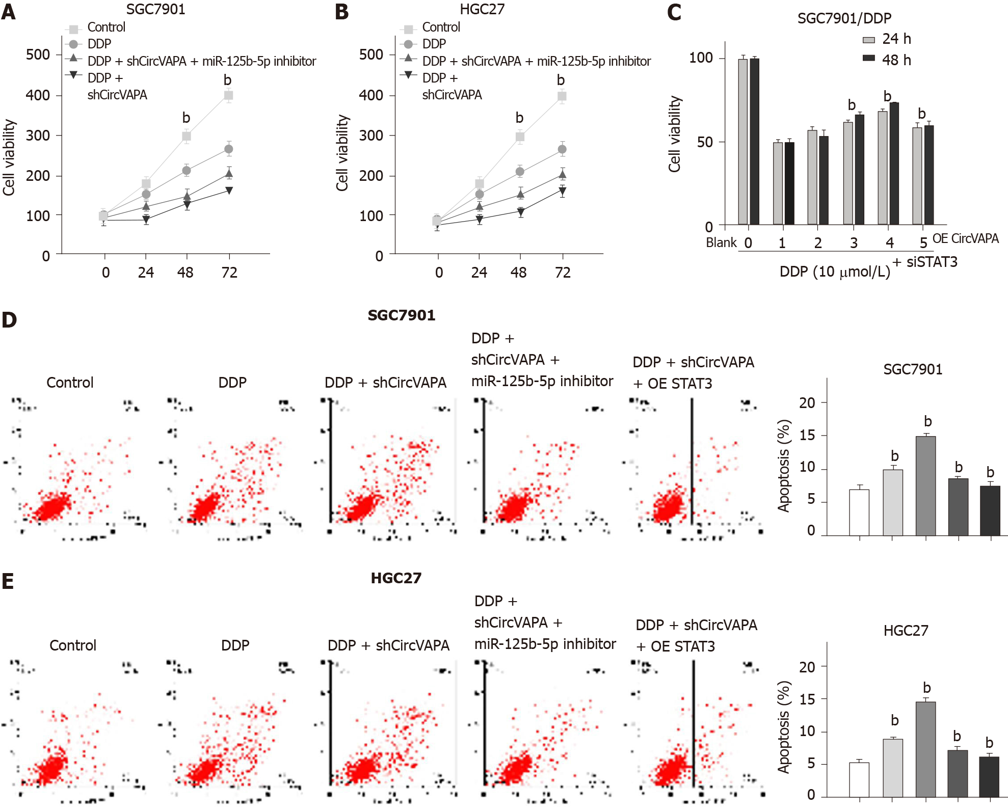
Figure 6 CircVAPA promotes chemotherapy drug resistance of gastric cancer cells by modulating miR-125b-5p/STAT3 axis.
A and B: SGC7901 and HGC27 cells were treated with cisplatin (DDP), DDP, and circVAPA shRNA, or co-treated with DDP, circVAPA shRNA, and miR-125b-5p inhibitor. Cell viability was measured by MTT assay; C: SGC7901/DDP cells were treated with DDP or co-treated with DDP and pcDNA3.1-circVAPA at the indicated dose, or co-treated with DDP, pcDNA3.1-circVAPA, and STAT3 siRNA. Cell viability was measured by MTT assay; D and E: SGC7901 and HGC27 cells were treated with DDP, DDP, and circVAPA shRNA, or co-treated with DDP, circVAPA shRNA, and miR-125b-5p inhibitor or pcDNA3.1-STAT3. Cell apoptosis was analyzed by flow cytometry. Data are presented as the mean ± SD. bP < 0.01. DDP: Cisplatin.
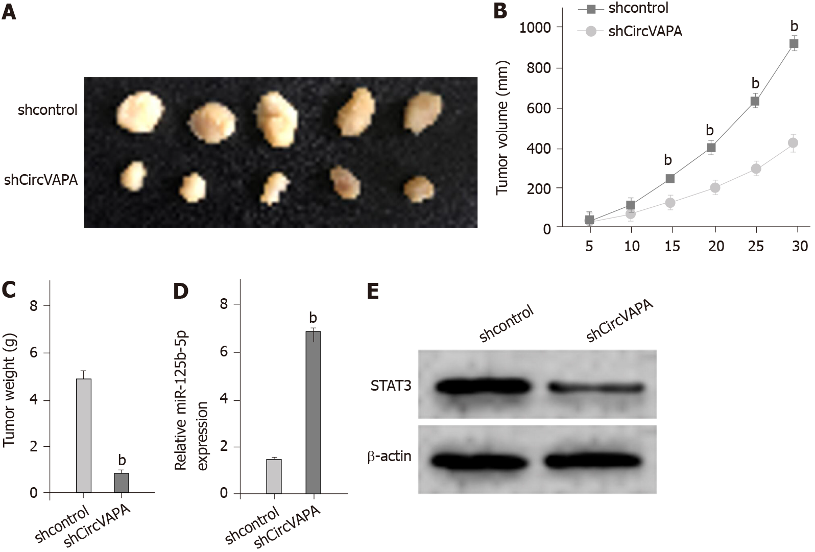
Figure 7 CircVAPA promotes tumor growth of chemoresistant gastric cancer cells in vivo.
A-D: The impact of circVAPA on tumor growth of melanoma cells in vivo was analyzed by nude mice tumorigenicity assay (n = 5). SGC7901/cisplatin cells were treated with control shRNA or circVAPA shRNA. Representative images of dissected tumors from nude mice are shown (A). The average tumor volume (B) and weight (C) were calculated and presented. The expression of miR-125b-5p was measured by qPCR in the tumor tissues of the mice (D); E: The expression of STAT3 was tested by Western blot analysis in the tumor tissues of the mice. Data are presented as the mean ± SD. bP < 0.01.
- Citation: Deng P, Sun M, Zhao WY, Hou B, Li K, Zhang T, Gu F. Circular RNA circVAPA promotes chemotherapy drug resistance in gastric cancer progression by regulating miR-125b-5p/STAT3 axis. World J Gastroenterol 2021; 27(6): 487-500
- URL: https://www.wjgnet.com/1007-9327/full/v27/i6/487.htm
- DOI: https://dx.doi.org/10.3748/wjg.v27.i6.487


