Copyright
©The Author(s) 2021.
World J Gastroenterol. Sep 28, 2021; 27(36): 6154-6160
Published online Sep 28, 2021. doi: 10.3748/wjg.v27.i36.6154
Published online Sep 28, 2021. doi: 10.3748/wjg.v27.i36.6154
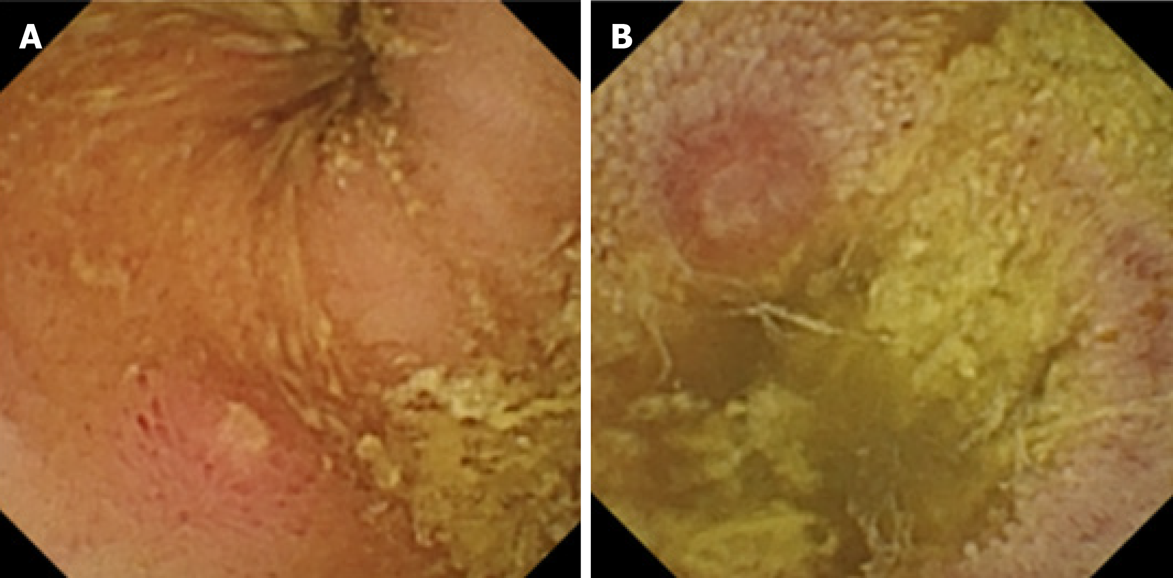
Figure 1 Capsule endoscopy with protruding lesion.
A: Capsule endoscopy with protruding lesion, with a depressed erosion at the tip suggestive of Meckel’s diverticulum; B: Capsule endoscopy with protruding lesion suggestive of Meckel’s diverticulum.
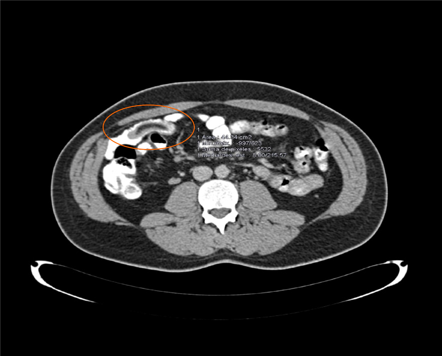
Figure 2 Abdominal computed tomography scan revealed a central area of fat attenuation surrounded by a thick collar of soft tissue attenuation suggestive of Meckel’s diverticulum.
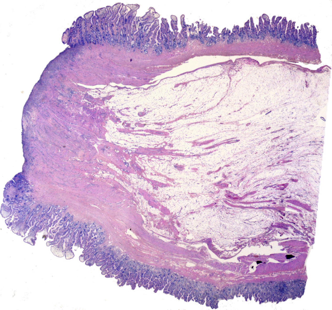
Figure 3 Low power histologic examination of a polypoid lesion lined by an intestinal type mucosa with a central ulcerated area.
No gastric or pancreatic heterotopic tissue can be found.
- Citation: El Hajra Martínez I, Calvo M, Martínez-Porras JL, Gomez-Pimpollo Garcia L, Rodriguez JL, Leon C, Calleja Panero JL. Inverted Meckel’s diverticulum diagnosed using capsule endoscopy: A case report. World J Gastroenterol 2021; 27(36): 6154-6160
- URL: https://www.wjgnet.com/1007-9327/full/v27/i36/6154.htm
- DOI: https://dx.doi.org/10.3748/wjg.v27.i36.6154


