Copyright
©The Author(s) 2021.
World J Gastroenterol. Sep 28, 2021; 27(36): 6079-6092
Published online Sep 28, 2021. doi: 10.3748/wjg.v27.i36.6079
Published online Sep 28, 2021. doi: 10.3748/wjg.v27.i36.6079
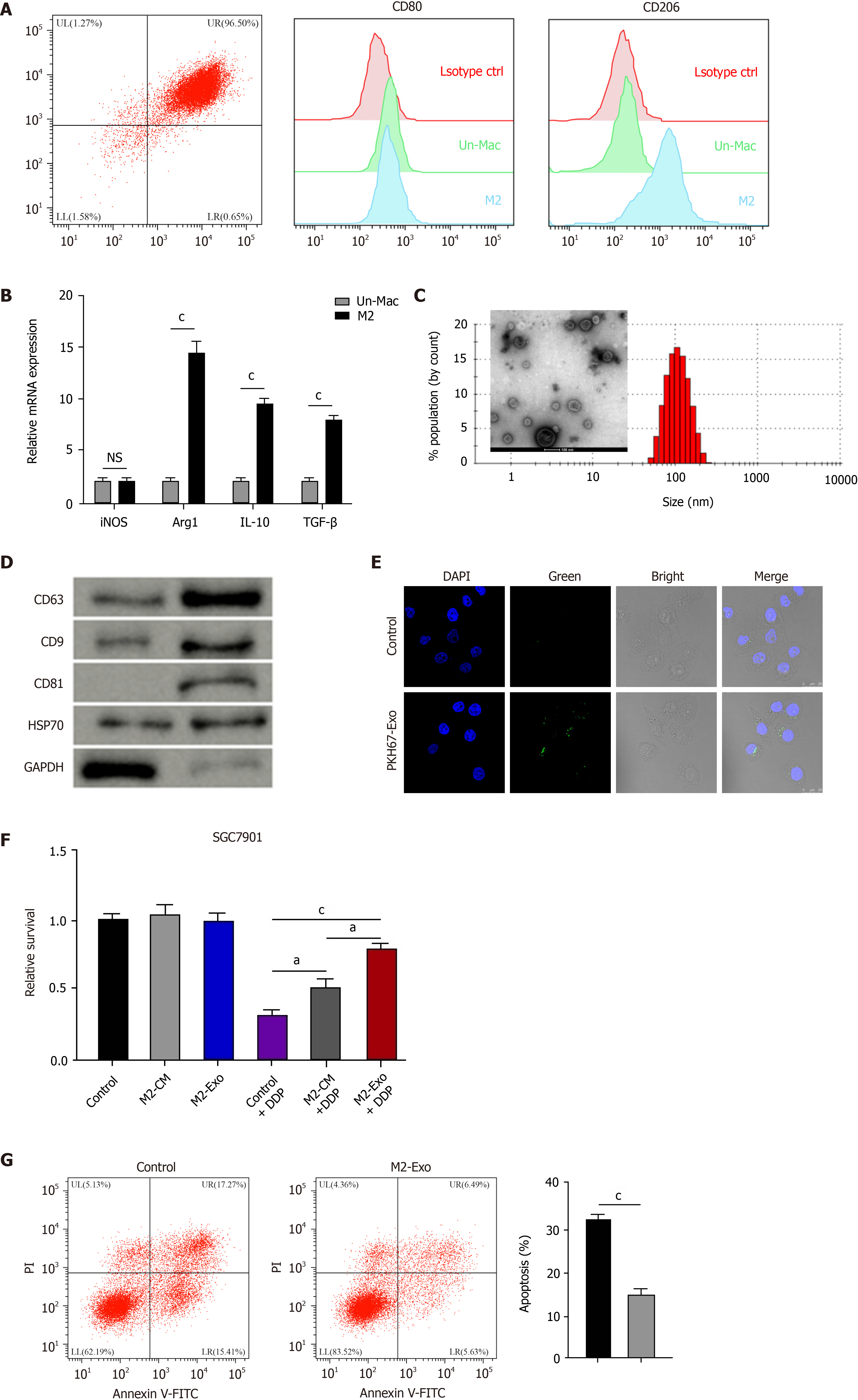
Figure 1 M2 polarized macrophages-derived exosomes can transfer in gastric cancer cells to enhance cisplatin resistance.
A and B: Flow cytometry for identifying the M2 polarized macrophages isolated from murine bone marrow induced with interleukin (IL)-13 and IL-4. The M2 specific marker CD206 was identified. Un-Mac represents inactivated macrophages and M2 represents macrophages activated by IL-13 and IL-4. The mRNA expression of iNOS, Arg1, IL-10, and TGF-β was detected by qPCR in Un-Mac and M2 macrophages; C: Identification of exosomes from M2 macrophages by transmission electron microscopy; D: Detection of expression of CD63, CD9, CD81, HSP70, and GAPDH by Western blot analysis; E: Detection of uptake of the PKH67-labelled M2 exosomes (M2-Exo) in SGC7901 cells; F: Analysis of cell viability by CCK-8 assay. M2 macrophages-derived conditioned medium and M2-Exo were used to treat SGC7901 cells exposed to cisplatin (DDP) for 48 h; G: Analysis of cell apoptosis by flow cytometry SGC7901 cells were co-treated with DDP and M2-Exo. Data shown are the mean ± SD. aP <0.05; cP < 0.001. DDP: Cisplatin; Un-Mac: Inactivated macrophages; M2-Exo: M2 exosomes.
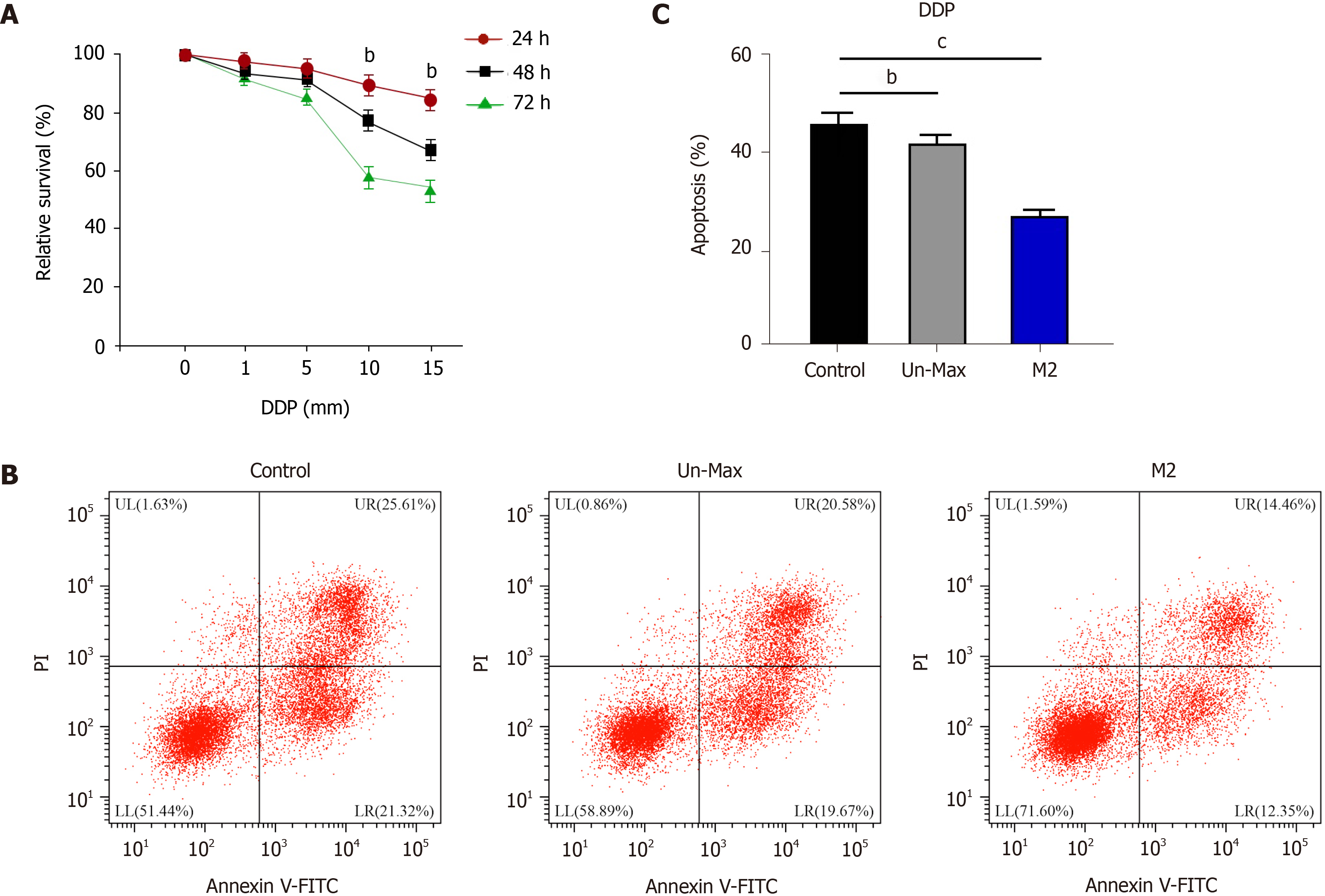
Figure 2 Co-culture of gastric cancer cells with M2 polarized macrophages promotes cisplatin resistance.
A: Detection of cell viability by CCK-8 assay in SGC7901 cells treated with cisplatin (DDP); B and C: Analysis of cell apoptosis by flow cytometry in SGC7901 cells co-cultured with inactivated macrophages and M2 macrophages exposed to DDP for 48 h. Data shown are the mean ± SD. bP < 0.01; cP <0.001. DDP: Cisplatin.
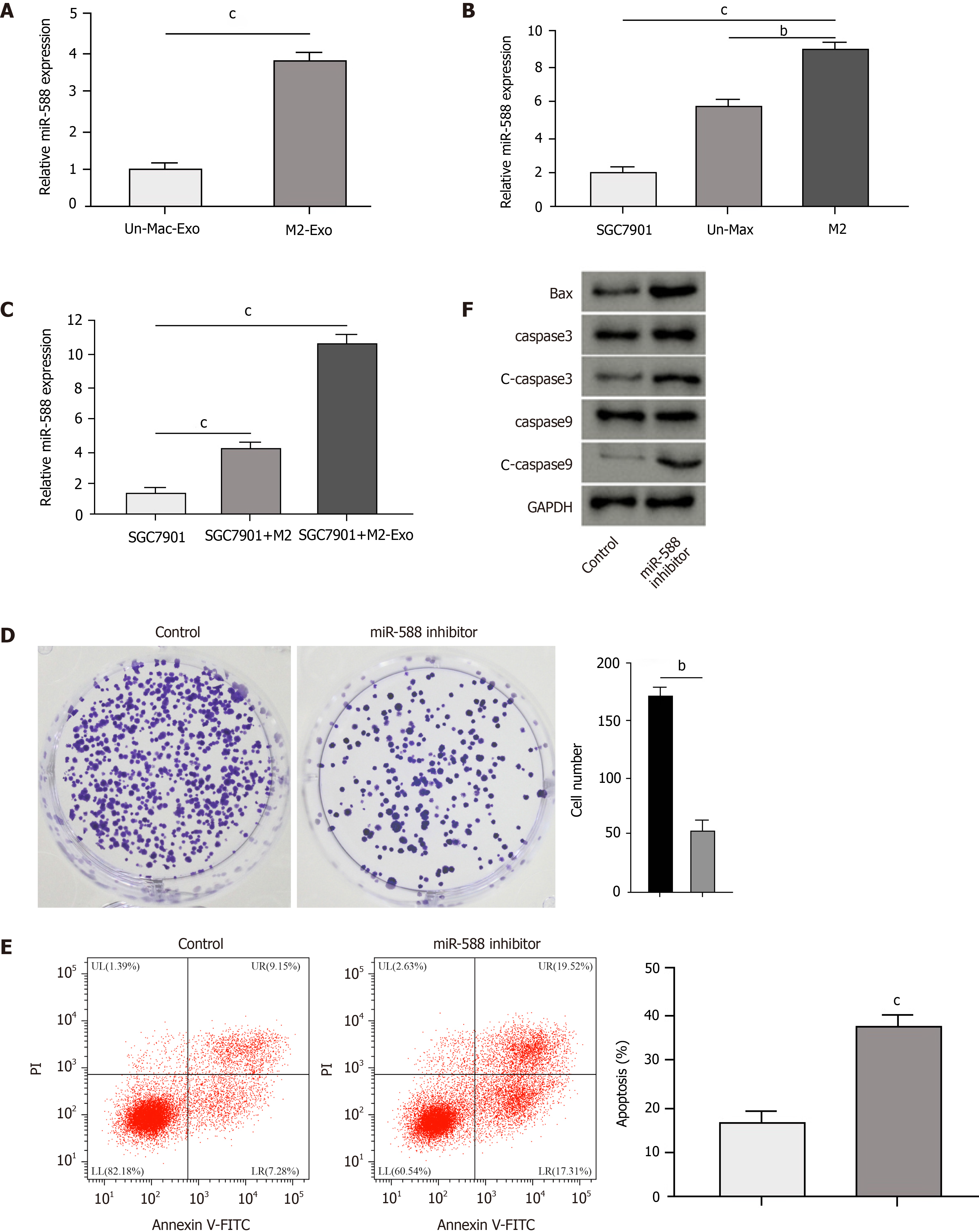
Figure 3 Exosomal miR-588 from M2 macrophages contributes to cisplatin resistance of gastric cancer cells.
A: Detection of expression of miR-588 by qPCR in the exosomes from M2 macrophages and inactivated macrophages; B: Detection of expression of miR-588 by qPCR in cisplatin (DDP)-resistant SGC7901 cells, M2 macrophages, and inactivated macrophages; C: Detection of expression of miR-588 by qPCR in DDP-resistant SGC7901 cells co-cultured with M2 macrophages or exosomes from M2 macrophages; D-F: Analysis of cell proliferation by colony formation assay (D), cell apoptosis by flow cytometry (E), and expression of Bax, capase-3, caspase-9, cleaved caspase-3 (c-caspase-3), and cleaved caspase 9 (c-caspase-9) by Western blot analysis (F) in DDP-resistant SGC7901 cells treated with miR-588 inhibitor. Data shown are the mean ± SD, bP < 0.01; cP < 0.001.
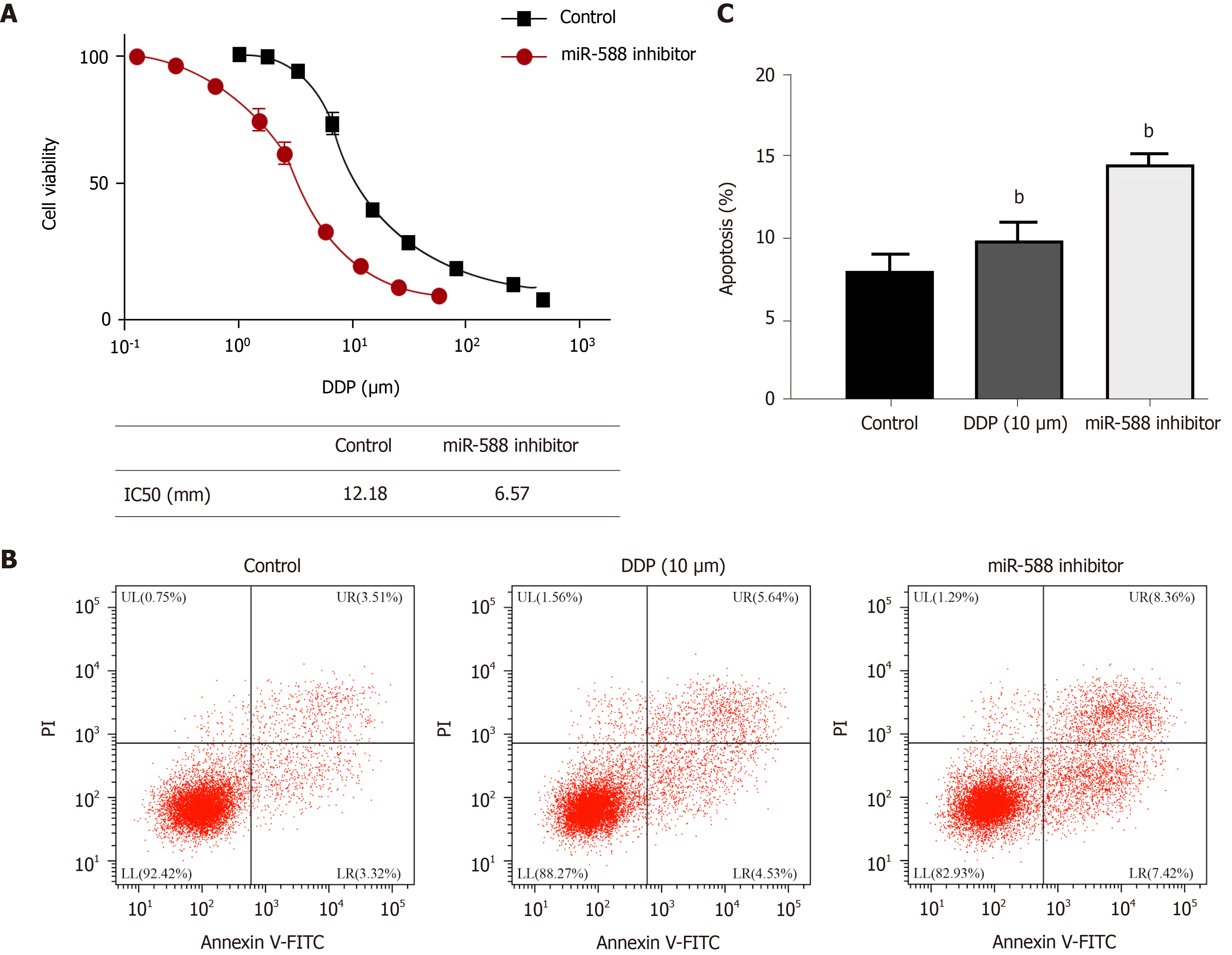
Figure 4 miR-588 contributes to cisplatin resistance of gastric cancer cells.
A: Detection of cell viability by CCK-8 assay in SGC7901 cells treated with miR-588 inhibitor and exposed to cisplatin (DDP) at the indicated doses; B and C: Detection of cell apoptosis by flow cytometry in DDP-resistant SGC7901 cells co-treated with DDP and miR-588 inhibitor. Data shown are the mean ± SD, bP < 0.01. DDP: Cisplatin.
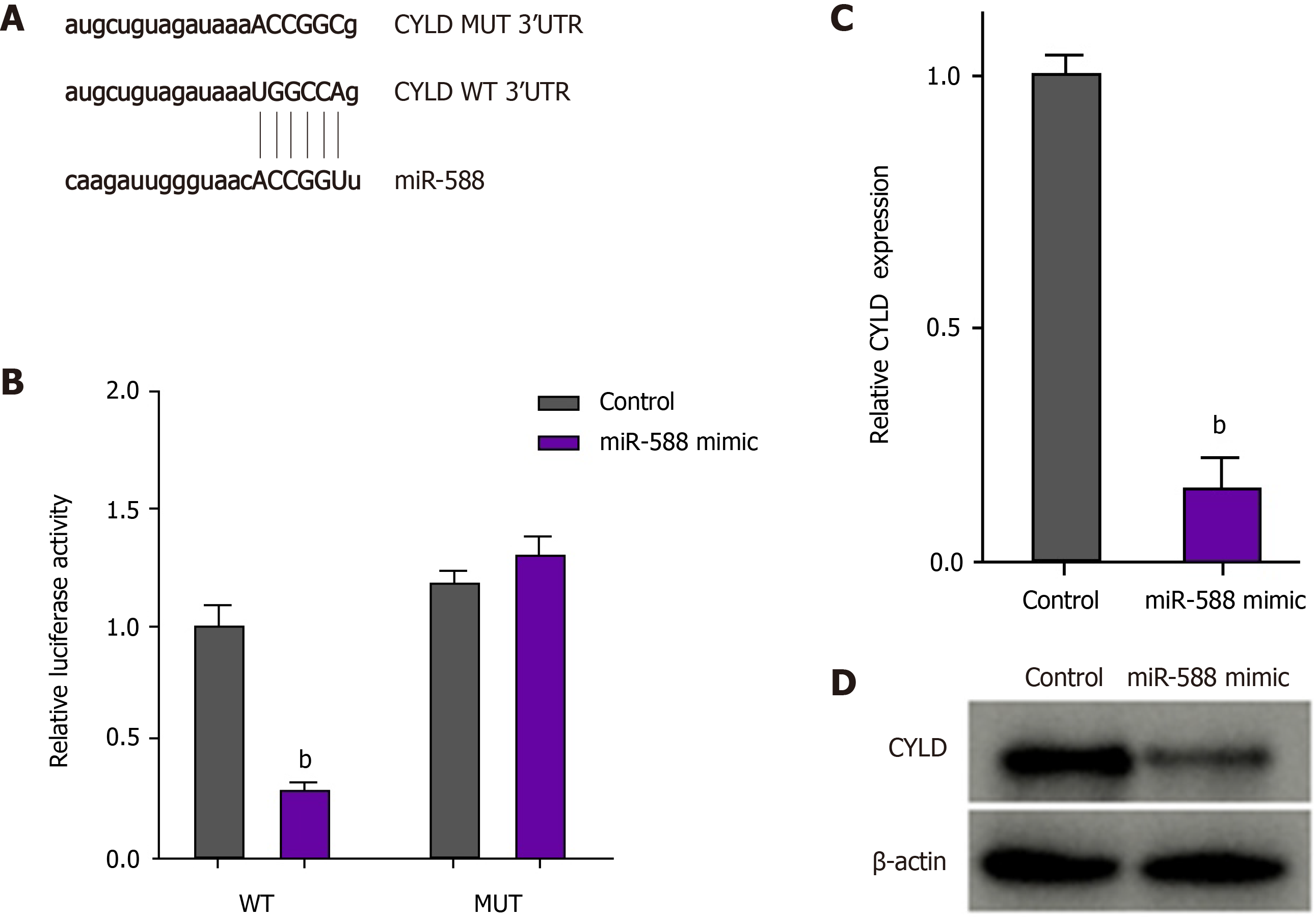
Figure 5 miR-588 can target cylindromatosis in gastric cancer cells.
A: The binding site of miR-588 and cylindromatosis (CYLD) was predicted in TargetScan database; B-D: After cisplatin (DDP)-resistant SGC7901 cells were treated with miR-588 mimic, the luciferase activity of CYLD was measured by luciferase reporter assays (B); the expression of CYLD was determined by qPCR (C); and the expression of CYLD was measured by Western blot analysis (D). Data shown are the mean ± SD. bP < 0.01. CYLD: Cylindromatosis.
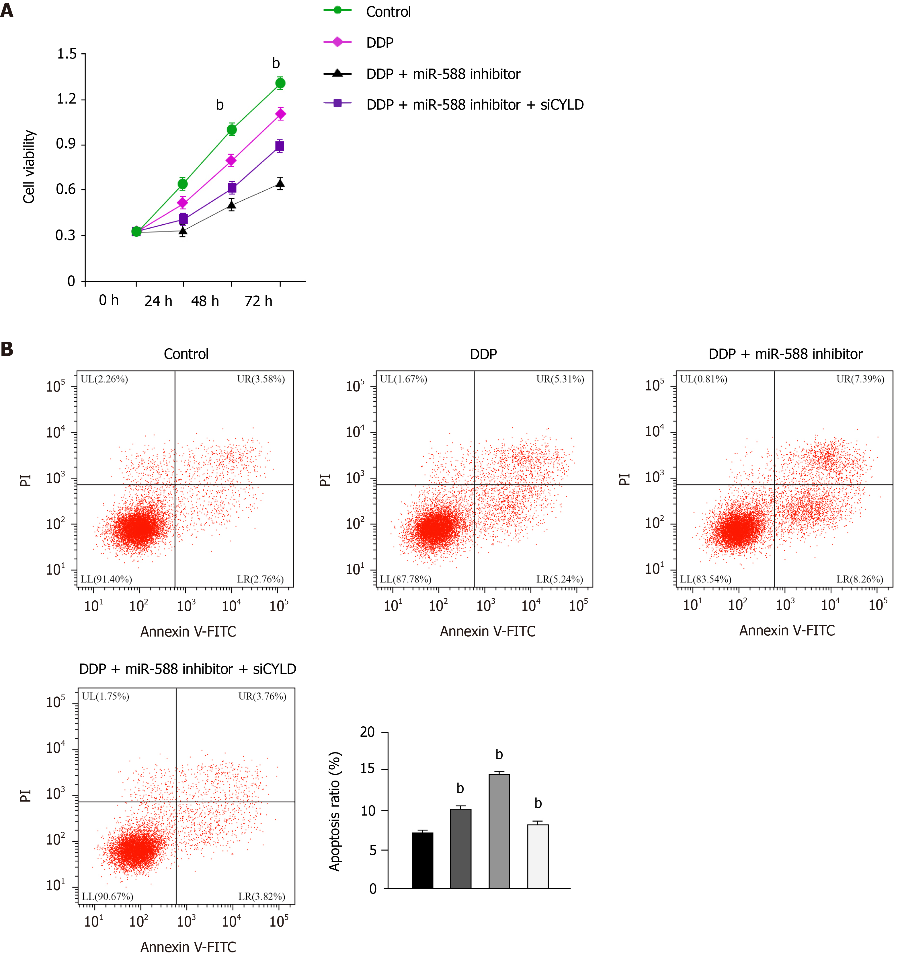
Figure 6 miR-588/cylindromatosis axis regulates cisplatin resistance of gastric cancer cells.
A: Cell viability detected by CCK-8 assay; B: Cell apoptosis analyzed by flow cytometry. The SGC7901 cells were treated with cisplatin (DDP), or co-treated with DDP and miR-588 inhibitor with or without cylindromatosis siRNA. Data shown are the mean ± SD. bP < 0.01. DDP: Cisplatin; CYLD: Cylindromatosis.
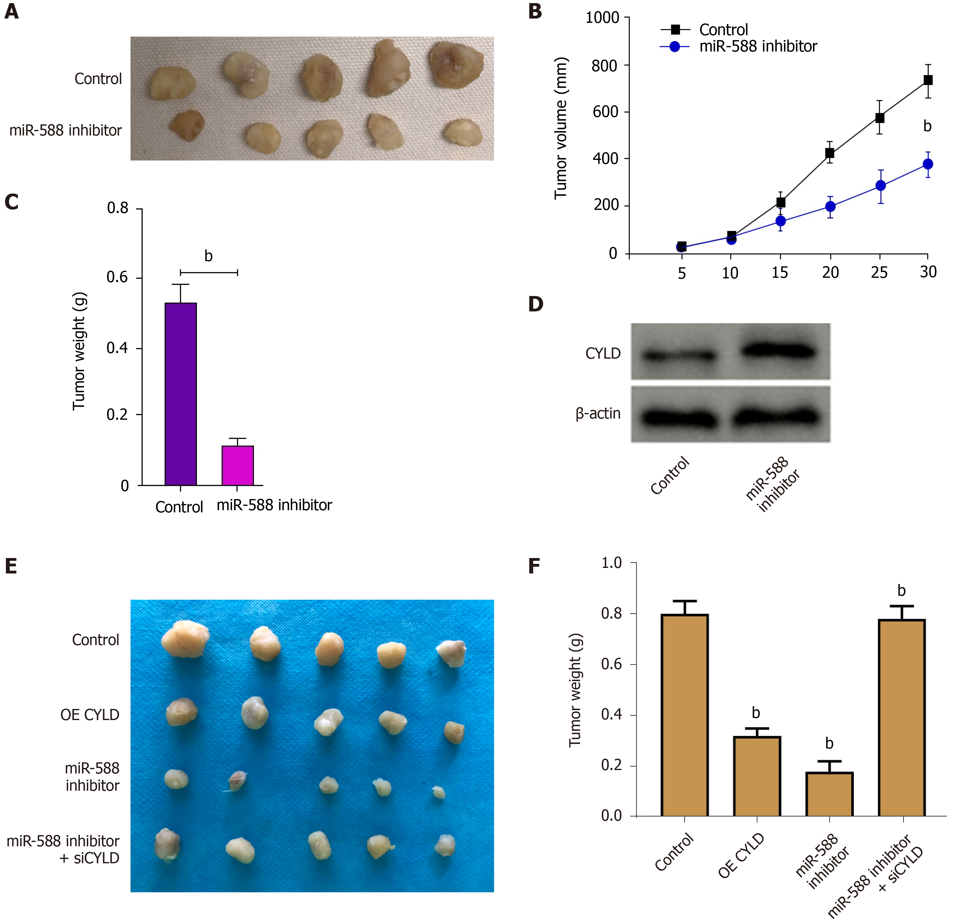
Figure 7 miR-588/cylindromatosis axis modulates cisplatin-resistant gastric cancer cell growth in vivo.
A-D: The nude mice were injected with SGC7901 cells treated with miR-588 inhibitor, the tumor tissues (A), tumor volume (B), and tumor weight (C) were measured, and the expression of cylindromatosis (CYLD) was detected by Western blot analysis in the tumor tissues (D); E and F: The nude mice were injected with SGC7901 cells treated with CYLD overexpressing plasmid or miR-588 inhibitor, or co-treated with miR-588 inhibitor and CYLD siRNA, and the tumor tissues (E) and tumor weight (F) were measured. Data shown are the mean ± SD (n = 5). bP < 0.01. CYLD: Cylindromatosis.
- Citation: Cui HY, Rong JS, Chen J, Guo J, Zhu JQ, Ruan M, Zuo RR, Zhang SS, Qi JM, Zhang BH. Exosomal microRNA-588 from M2 polarized macrophages contributes to cisplatin resistance of gastric cancer cells. World J Gastroenterol 2021; 27(36): 6079-6092
- URL: https://www.wjgnet.com/1007-9327/full/v27/i36/6079.htm
- DOI: https://dx.doi.org/10.3748/wjg.v27.i36.6079


