Copyright
©The Author(s) 2021.
World J Gastroenterol. Jul 21, 2021; 27(27): 4252-4275
Published online Jul 21, 2021. doi: 10.3748/wjg.v27.i27.4252
Published online Jul 21, 2021. doi: 10.3748/wjg.v27.i27.4252
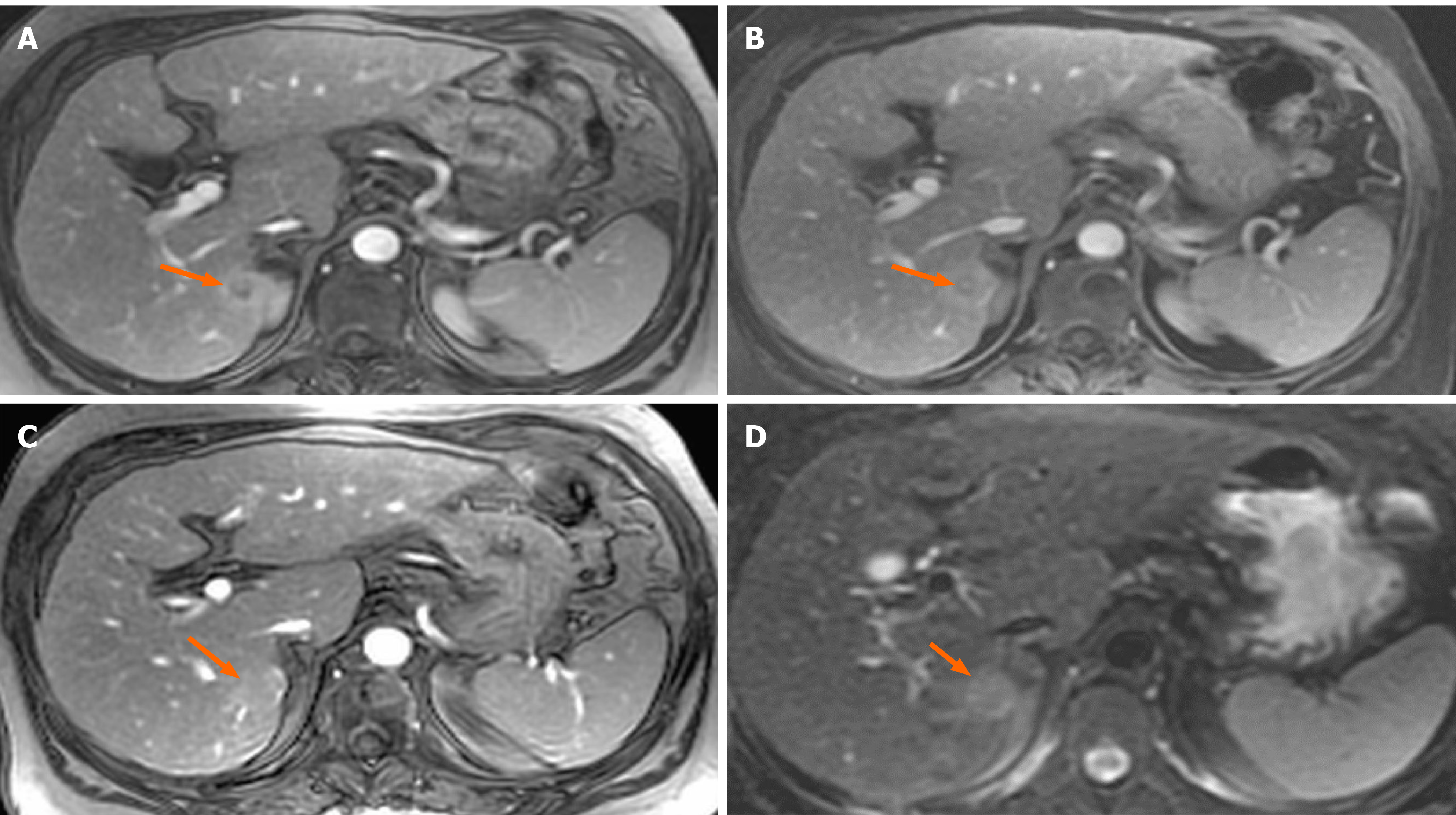
Figure 1 A small (1.
8 cm) liver nodule with arterial enhancement is discovered in the left hepatic lobe during magnetic resonance imaging. A: T1, arterial phase; B: T1 portal phase; C: T1 delayed phase; D: T2.
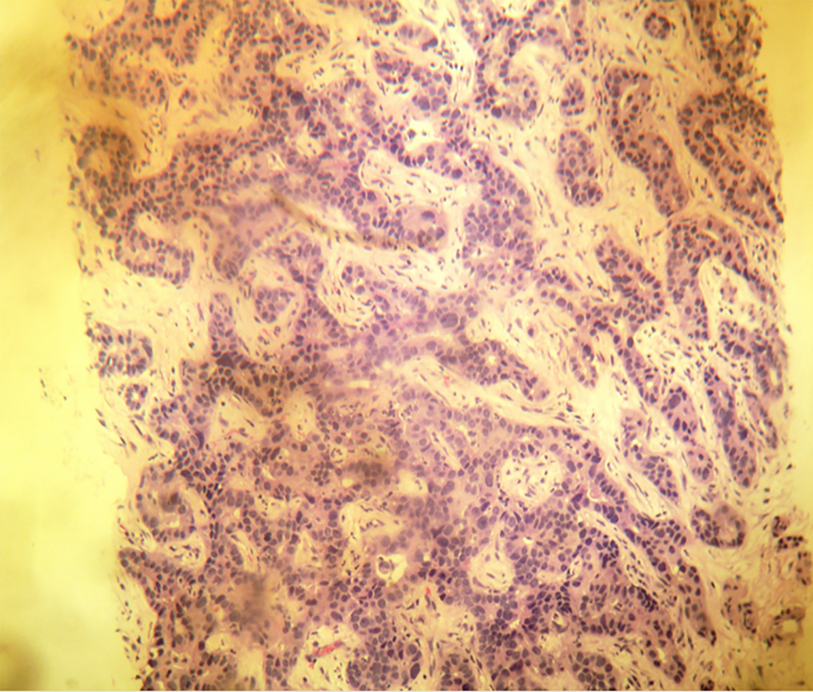
Figure 2 Histological findings typical of cholangiocarcinoma.
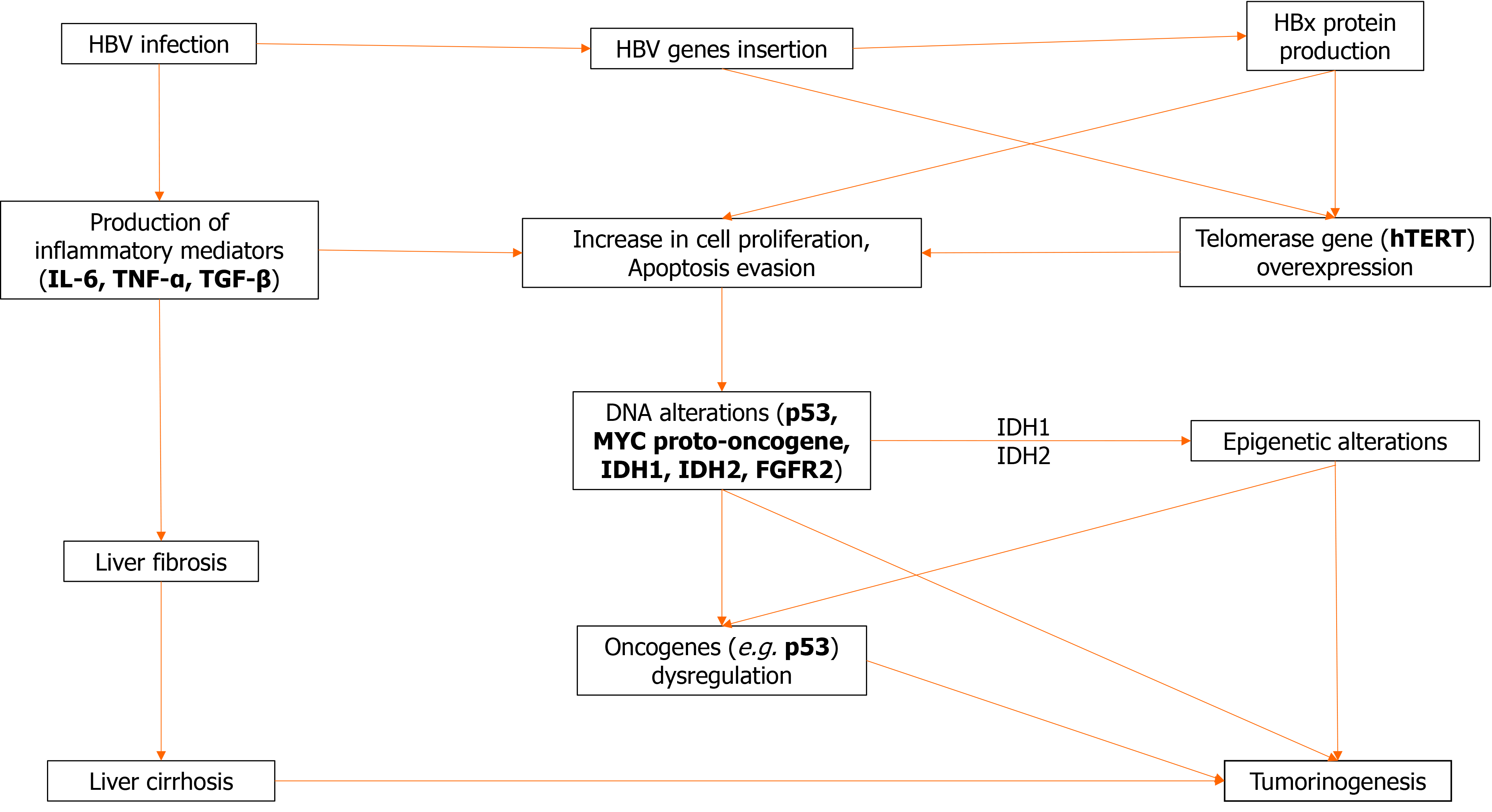
Figure 3 Pathological mechanisms that lead to tumorigenesis in patients with hepatitis B virus infection.
FGFR: Fibroblast growth factor receptor; HBV: Hepatitis B virus; IDH: Isocitrate dehydrogenase; IL-6: Interleukin 6; TGF-β: Transforming growth factor β; TNF-α: Tumor necrosis factor α.
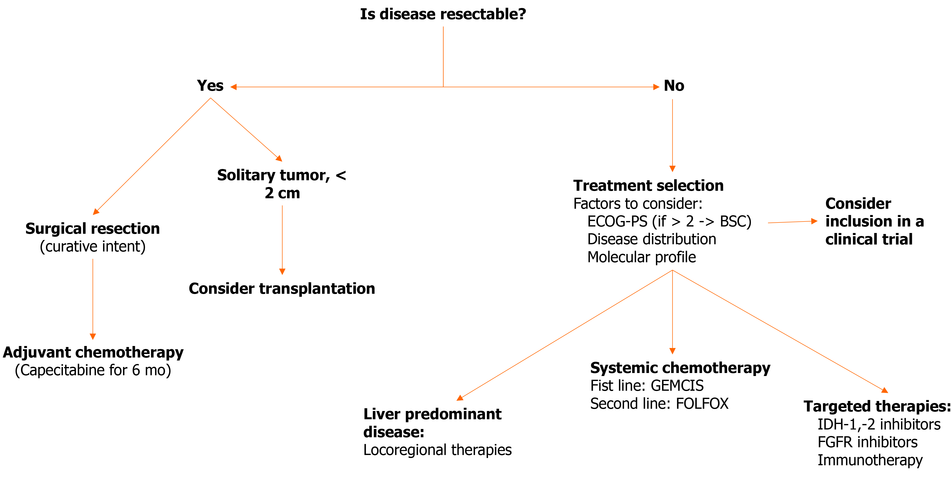
Figure 4 Therapeutic algorithm for hepatitis B virus-associated intrahepatic cholangiocarcinoma.
BSC: Best supportive care; ECOG-PS: Eastern Cooperative Oncology Group-Performance Status; FGFR: Fibroblast growth factor receptor; FOLFOX: Folinic acid, Fluorouracil and Oxaliplatin combination; GEMCIS: Gemcitabine and cisplatin combination; IDH: Isocitrate dehydrogenase.
- Citation: Fragkou N, Sideras L, Panas P, Emmanouilides C, Sinakos E. Update on the association of hepatitis B with intrahepatic cholangiocarcinoma: Is there new evidence? World J Gastroenterol 2021; 27(27): 4252-4275
- URL: https://www.wjgnet.com/1007-9327/full/v27/i27/4252.htm
- DOI: https://dx.doi.org/10.3748/wjg.v27.i27.4252


