Copyright
©The Author(s) 2021.
World J Gastroenterol. Jun 21, 2021; 27(23): 3327-3341
Published online Jun 21, 2021. doi: 10.3748/wjg.v27.i23.3327
Published online Jun 21, 2021. doi: 10.3748/wjg.v27.i23.3327
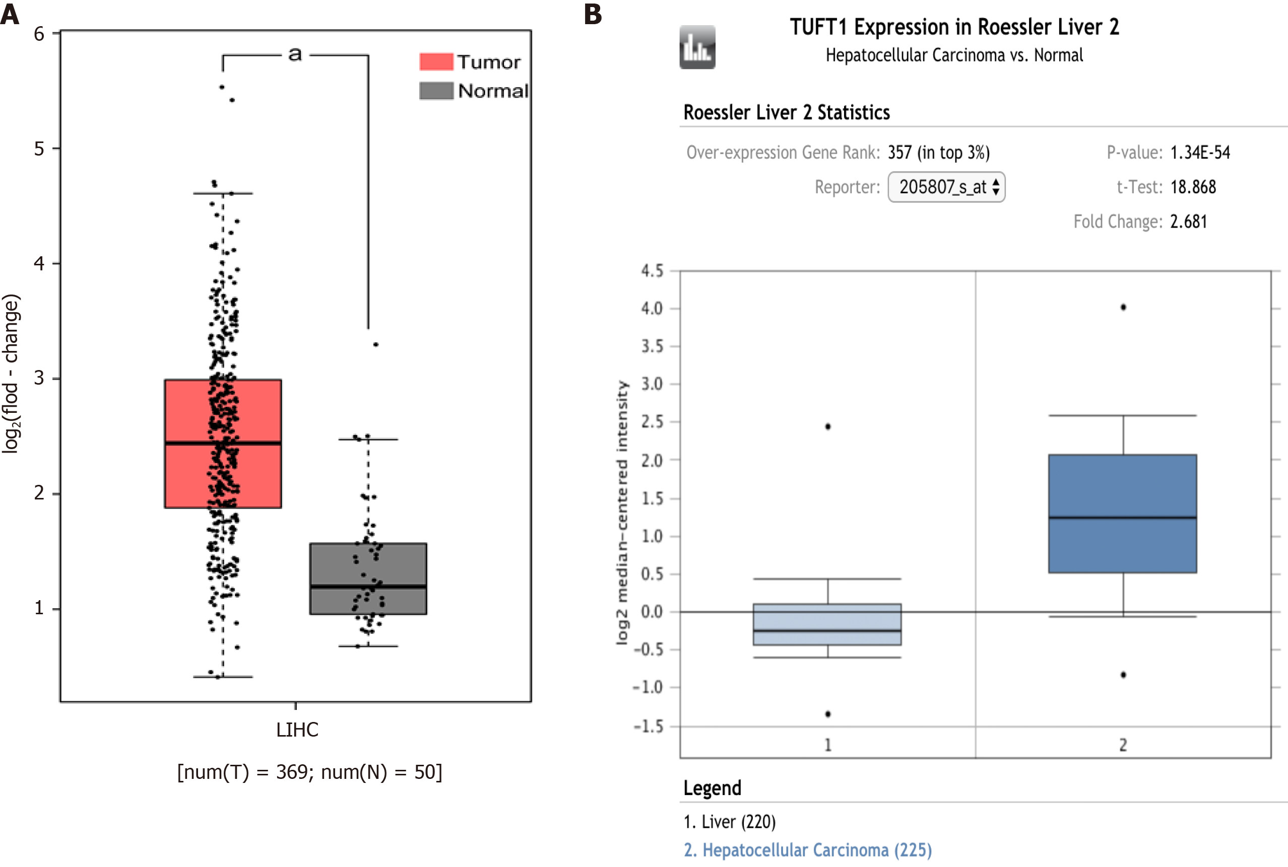
Figure 1 Comparative analysis of tuftelin 1 messenger RNA between hepatocellular carcinoma and normal livers based on bioinformatics databases.
A: The expression of tuftelin 1 (TUFT1) messenger RNA in the cancerous tissues of hepatocellular carcinoma (HCC) patients (n = 369, red, aP < 0.05) and normal liver tissues (n = 50, grey) from the Cancer Genome Atlas database; B: The expression of TUFT1 messenger RNA in the cancerous tissues of HCC patients (n = 225, dark blue, aP < 0.05) and normal liver tissues (n = 220, light blue) from the Oncomine database. The horizontal line represents the median of the two groups. Vertical bars indicate the range of data. LIHC: Liver hepatocellular carcinoma.
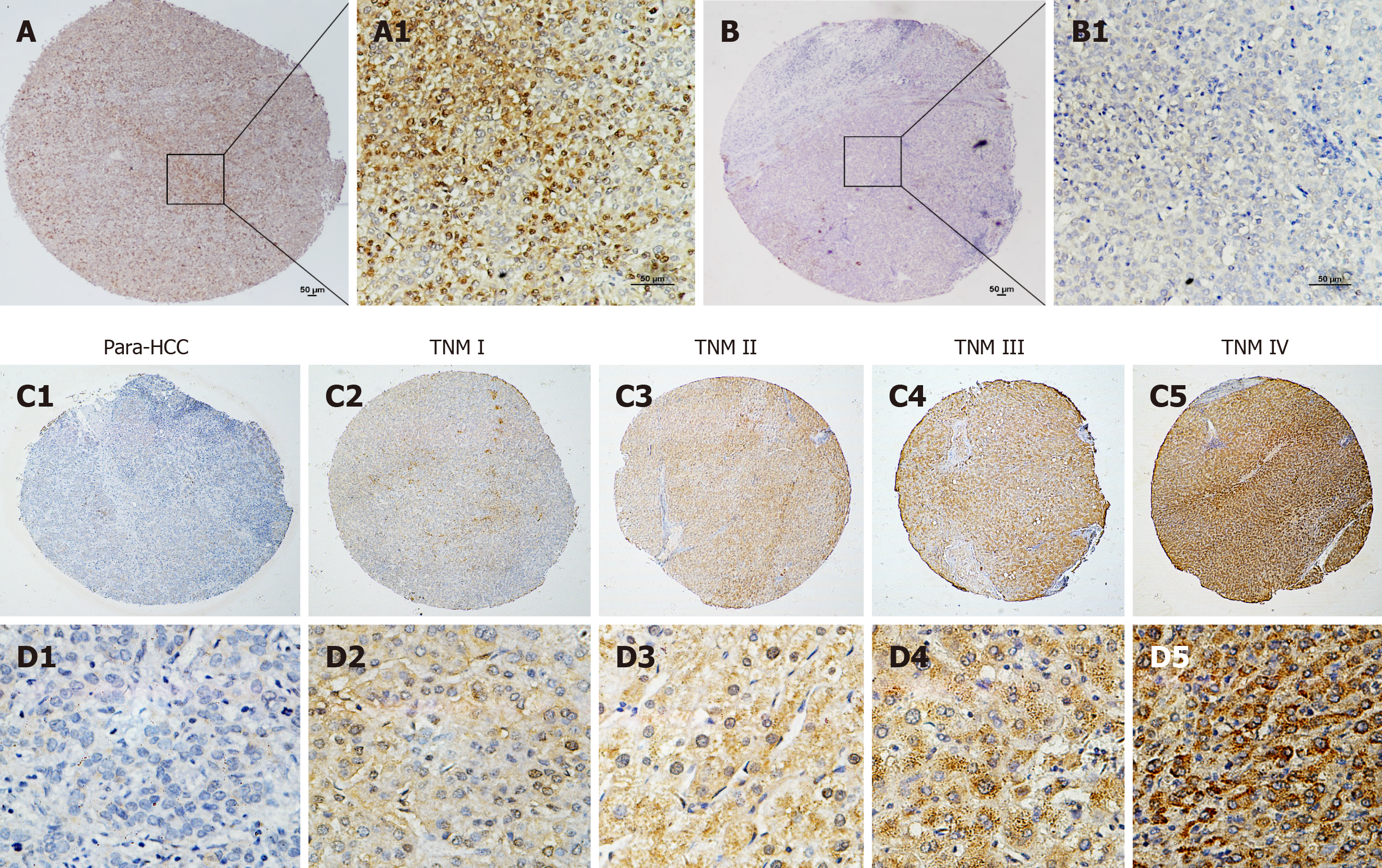
Figure 2 Tuftelin 1 expression with clinical staging of hepatocellular carcinoma.
A, A1: Hepatocellular carcinoma (HCC) tissue with strongly positive staining; B, B1: Adjacent non-HCC tissue with negative staining, original magnifications of × 40 (scale bar: 50 μm) in A and B, and × 200 (scale bar: 50 μm) in A1 and B1; C, D: Tuftelin 1 (TUFT1) expression in different HCC staging; C1, D1: Low TUFT1 expression in the non-HCC tissues; C2-C5: The brown staining of TUFT1 gradually increases in the HCC tissues from stage I to IV (original magnification × 40); D2-D5: The staining of TUFT1 in the non-HCC tissues from stage I to IV (original magnification × 400). TNM: Tumor-node-metastasis.
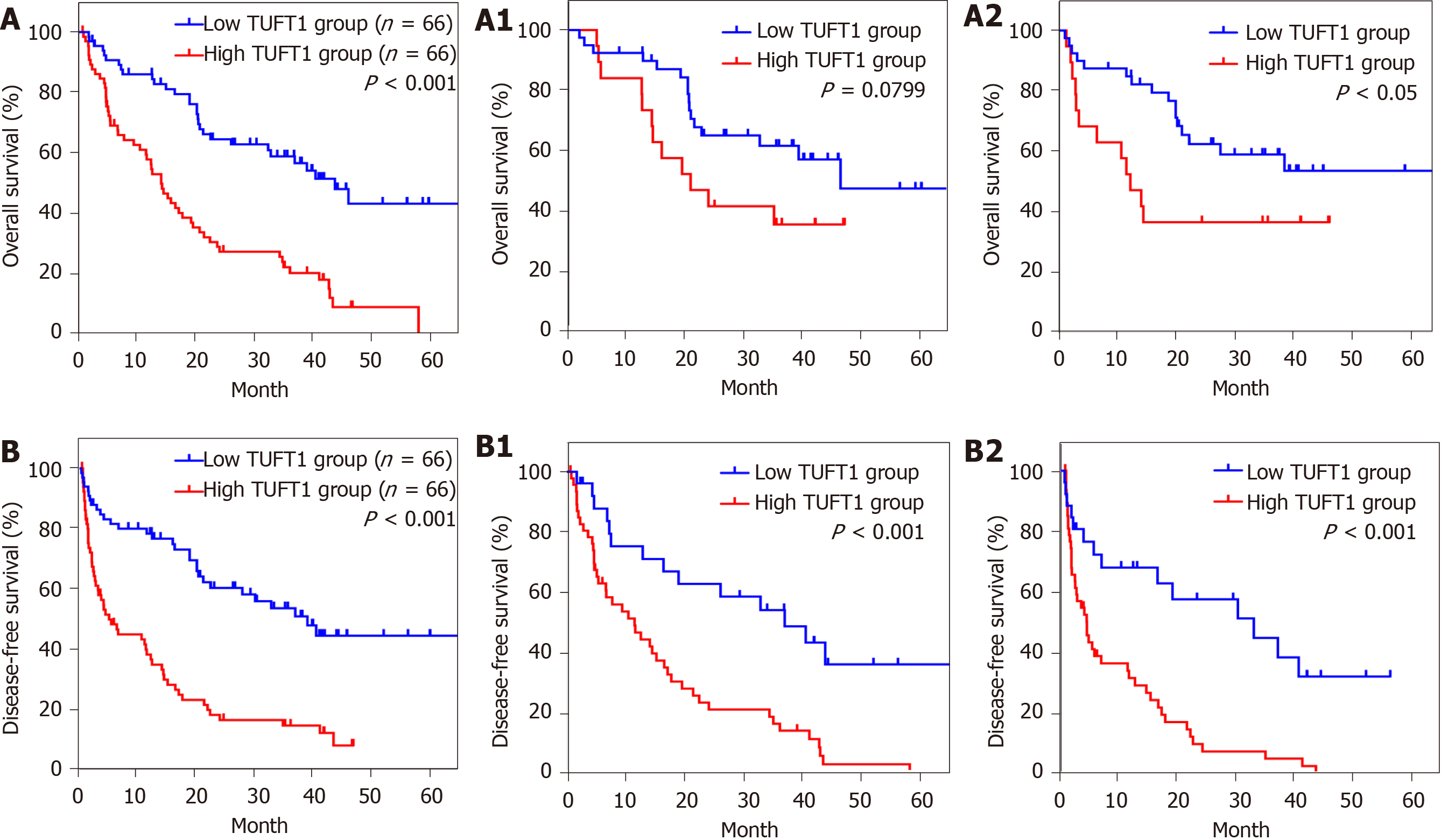
Figure 3 Relationship between tuftelin 1 expression and prognosis of hepatocellular carcinoma.
A: Kaplan-Meier analysis was performed to compare the overall survival (OS) between hepatocellular carcinoma (HCC) with high (n = 66) and low (n = 66) TUFT1; A1: The high (n = 19) and low (n = 40) tuftelin 1 (TUFT1) with OS at early HCC; A2: The high (n = 47) and low (n = 26) TUFT1 with OS at advanced HCC; B: Kaplan-Meier analysis was performed to compare the disease-free survival (DFS) between HCC with high (n = 66) and low (n = 66) TUFT1; B1: The high (n = 19) and low (n = 40) TUFT1 with DFS at early HCC; B2: The high (n = 47) and low (n = 26) TUFT1 with DFS at advanced HCC.
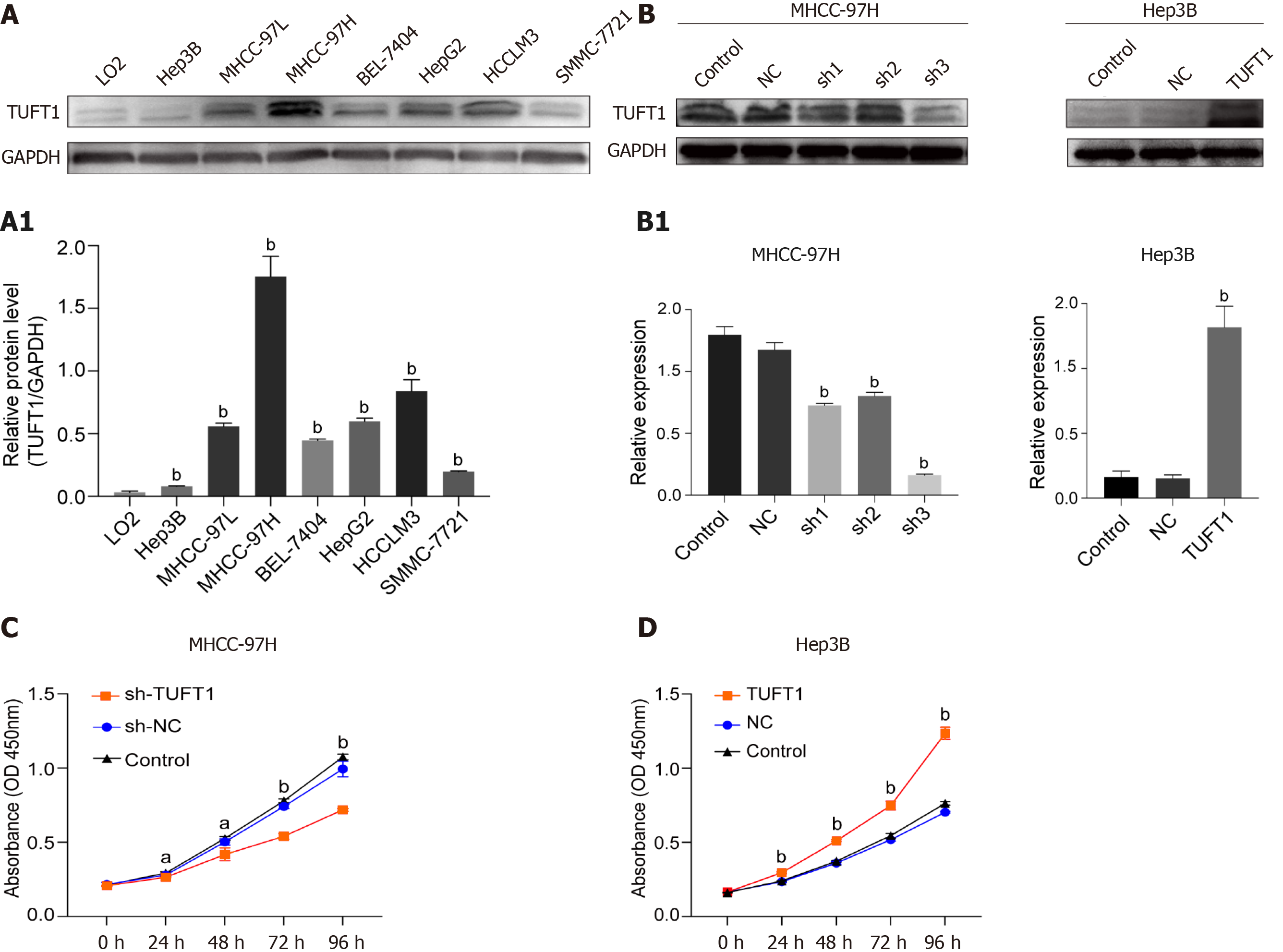
Figure 4 Expression, interfering of tuftelin 1 with proliferation of hepatocellular carcinoma cells.
A: Expressions of tuftelin 1 (TUFT1) among different hepatocellular carcinoma (HCC) cell lines were detected by Western blotting, and the expressing levels of all HCC cells were significantly higher than that of the control LO2 cells (bP < 0.001); A1: The relative ratio from TUFT1 to glyceraldehyde 3-phosphate dehydrogenase with the MHCC-97H cells showed the strongest TUFT1 expression, and the Hep3B cells showed the lowest TUFT1 expression; B: The MHCC-97H cells and interfering with TUFT1 short hairpin RNA (sh-RNA)1-3 (left); the TUFT1 was over-expressed after the Hep3B cells transfected with the constructed pEX-4 (pGCMV/MCS/T2A/EGFP/Neo) plasmid (Right); B1: The TUFT1 expression was significantly inhibited by the TUFT1-shRNA transfection (bP < 0.001, left); the markedly increasing TUFT1 Level was compared with the control cells (bP < 0.001, right); C: The TUFT1 activation interfering by TUFT1- shRNA3 significantly inhibited the proliferation of MHCC-97H cells (aP < 0.05, bP < 0.001); D: the over-expression of TUFT1 significantly enhanced the proliferation of the Hep3B cells (bP < 0.001). Data are presented as mean ± standard error of the mean of at least three independent experiments. NC: Blank control; sh-NC: Negative sh-RNA for tuftelin 1 mRNA.
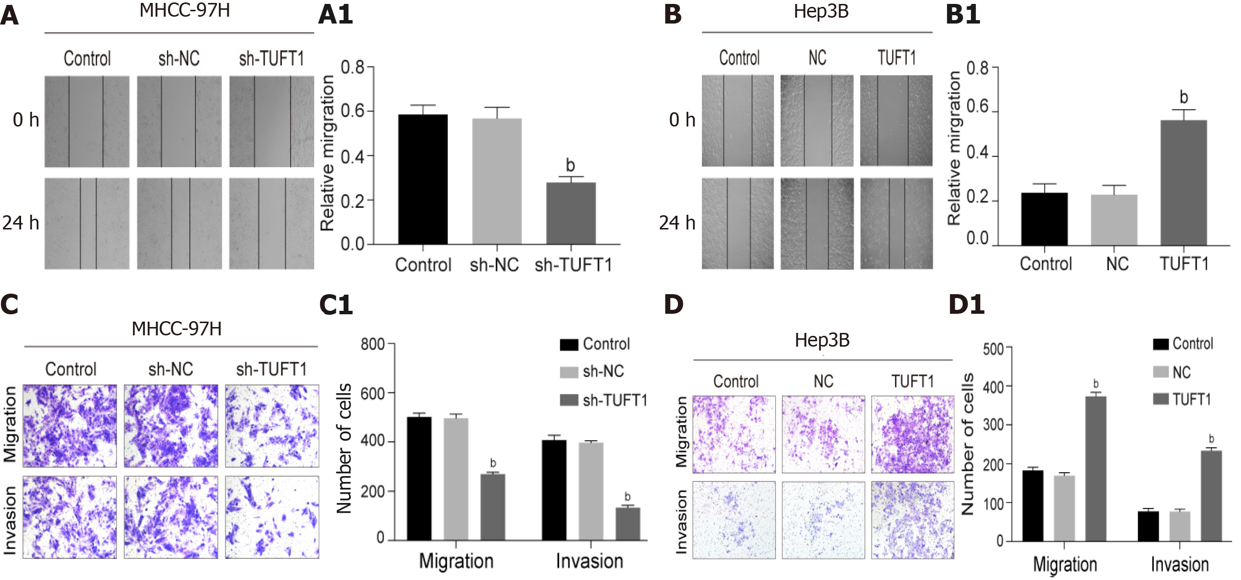
Figure 5 Effects of tuftelin 1 on healing, invasion, and migration of hepatocellular carcinoma cells.
A: The scratch test of the MHCC-97H cells, the cell healing ability was decreasing after the high tuftelin 1 (TUFT1) cells were transfected with the TUFT1-shRNA3 plasmid, and A1, the relative cell healing ability was significantly inhibited compared with the control or negative sh-RNA for tuftelin 1 mRNA (sh-NC) cells (bP < 0.001); B: The scratch test of the Hep3B cells, the cell healing ability was increased after the cells were transfected with the over-expressing TUFT1 plasmid; B1, the relative cell healing ability was significantly improved compared with the control or blank control (NC) cells (bP < 0.001); C: The migration and invasion abilities of the MHCC-97H cells were analyzed by the transwell test; C1: The histograms of the cell migration and invasion abilities among the different groups (bP < 0.001): D: The migration and invasion abilities of the Hep3B cells were analyzed by the transwell test; D1: The histograms of the cell migration and invasion abilities among the different groups (bP < 0.001). Data are presented as mean ± standard error of the mean of at least three independent experiments. sh-RNA: sh-RNA3 for tuftelin 1 mRNA.
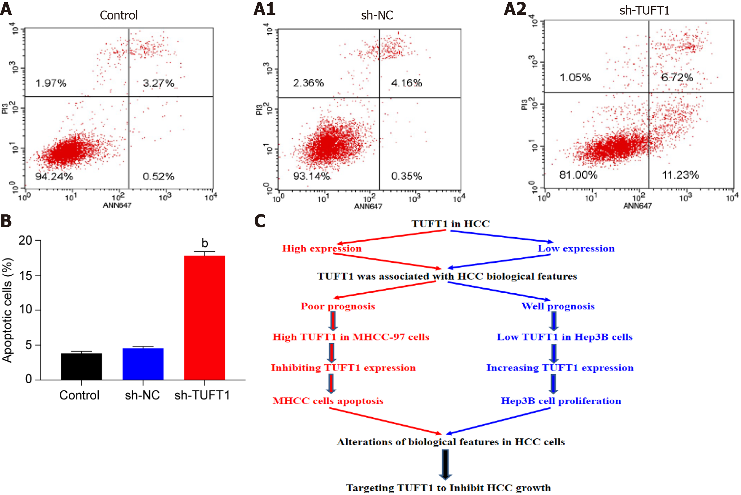
Figure 6 Possible mechanism of tuftelin 1 expression in hepatocellular carcinoma cells progression.
A: The MHCC-97H cells were transfected with the tuftelin 1 (TUFT1)-short hairpin RNA (sh-RNA)3 plasmid, and the apoptotic cells in the sh-TUFT1 group (A2) were increased more than those in the blank control (NC) (A) or negative sh-RNA for tuftelin 1 mRNA (sh-NC) group (A1); B: The histograms of the apoptotic cells among the different groups, the rate of the MHCC-97H cell apoptosis in the sh-TUFT1 group was significantly higher (bP < 0.001) than those in the NC or sh-NC group. Data are presented as mean ± standard error of the mean of at least three independent experiments; C: Speculation of possible mechanisms of TUFT1 promotion of the progression of hepatocellular carcinoma (HCC). Aberrant TUFT1 activation promoted the proliferation and metastasis via anti-apoptosis of HCC cells.
- Citation: Wu MN, Zheng WJ, Ye WX, Wang L, Chen Y, Yang J, Yao DF, Yao M. Oncogenic tuftelin 1 as a potential molecular-targeted for inhibiting hepatocellular carcinoma growth. World J Gastroenterol 2021; 27(23): 3327-3341
- URL: https://www.wjgnet.com/1007-9327/full/v27/i23/3327.htm
- DOI: https://dx.doi.org/10.3748/wjg.v27.i23.3327


