Copyright
©The Author(s) 2021.
World J Gastroenterol. Jun 14, 2021; 27(22): 3037-3049
Published online Jun 14, 2021. doi: 10.3748/wjg.v27.i22.3037
Published online Jun 14, 2021. doi: 10.3748/wjg.v27.i22.3037
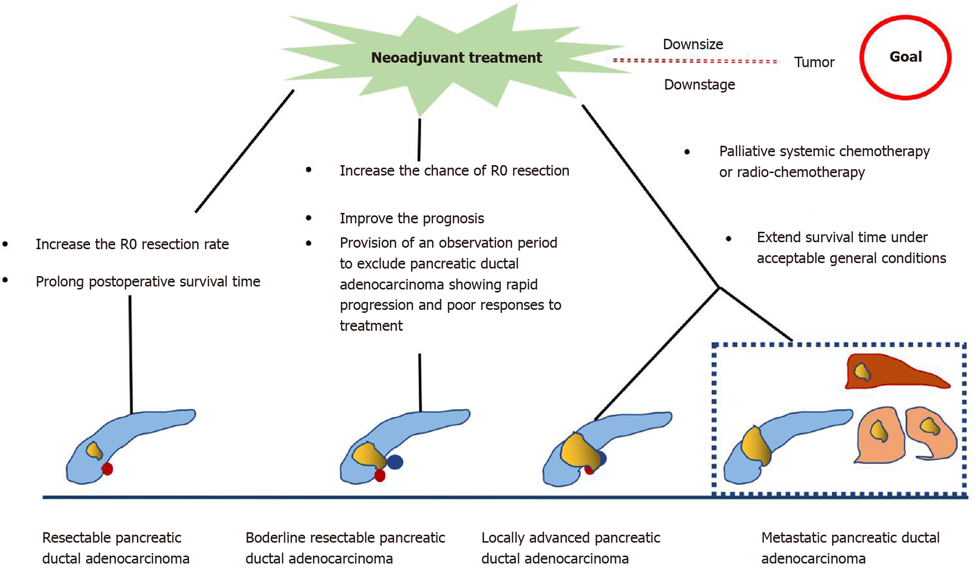
Figure 1
Role of neoadjuvant treatment for different types of pancreatic ductal adenocarcinoma.
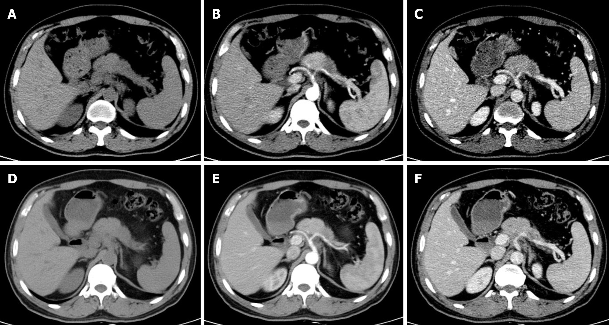
Figure 2 Response assessment with contrast-enhanced computed tomography after neoadjuvant treatment.
A-C: A 55-year-old man with a 2.4 cm tumor in the pancreas with non-uniform low density (A), showing no obvious enhancement relative to the surrounding pancreatic parenchyma in the arterial phase (B), without hyperenhancement or distinct ‘wash-out’ appearance in the portal venous phase (C); D-F: After 20 d of neoadjuvant treatment (FOLFOX), tumor size was reduced to 2.1 cm, with low enhancement relative to the surrounding pancreatic parenchyma on contrast-enhanced computed tomography images.
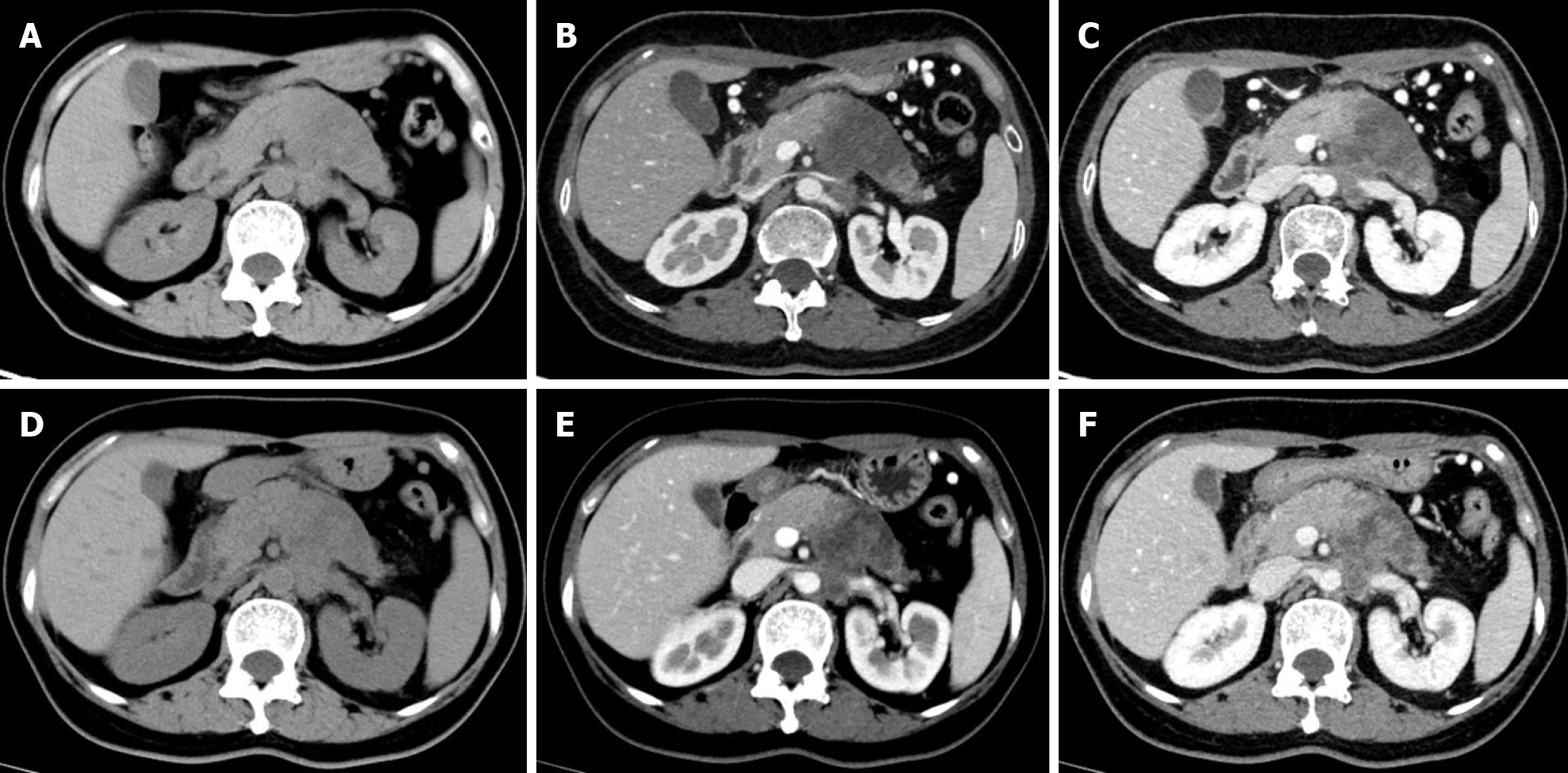
Figure 3 Response assessment with contrast-enhanced computed tomography after neoadjuvant therapy.
A-C: A 58-year-old woman with a 5.6 cm × 4.2 cm tumor in the pancreatic body and tail with non-uniform low-density (A), showing slightly enhancement relative to the surrounding parenchyma in the arterial phase (B), with invasion of the left renal vein as seen in the portal venous phase (C); D-F: After 2.5 mo of neoadjuvant treatment (nab-paclitaxel combined with gemcitabine), the tumor size was reduced to 4.7 cm × 4.2 cm (D), small patchy enhancement was seen in the original lesion (E), and the degree of invasion of the left renal vein was reduced (F).
- Citation: Zhang Y, Huang ZX, Song B. Role of imaging in evaluating the response after neoadjuvant treatment for pancreatic ductal adenocarcinoma. World J Gastroenterol 2021; 27(22): 3037-3049
- URL: https://www.wjgnet.com/1007-9327/full/v27/i22/3037.htm
- DOI: https://dx.doi.org/10.3748/wjg.v27.i22.3037


