Copyright
©The Author(s) 2021.
World J Gastroenterol. May 7, 2021; 27(17): 2015-2024
Published online May 7, 2021. doi: 10.3748/wjg.v27.i17.2015
Published online May 7, 2021. doi: 10.3748/wjg.v27.i17.2015
Figure 1 Screening process for patients with liver cancer.
HCC: Hepatocellular carcinoma; CT: Computed tomography; MVI: Microvascular invasion.
Figure 2 Specific performance of two-trait predictor of venous invasion and radiogenomic invasion.
A and B: The discriminant process of two-trait predictor of venous invasion (TTPVI) (A) and radiogenomic invasion (RVI) (B); C: Negative intratumoral arteries: Negative TTPVI and RVI; D: Positive intratumoral arteries and peritumoral low density: Negative TTPVI and PVI; E: Positive intratumoral arteries, negative peritumoral low-density shadow, and positive tumor-liver differences: Positive TTPVI and negative RVI; F: Positive intratumoral arteries, negative peritumoral low density, and negative tumor-liver differences: Positive TTPVI and RVI. TTPVI: Two-trait predictor of venous invasion; RVI: Radiogenomic invasion.
Figure 3 Selection of radiomic features using the least absolute shrinkage and selection operator-logistic regression model.
A: Coefficient profile of 158 radiomics features against the area under the curve; B: Cross-validation curve. Red dotted vertical lines are drawn at the optimal log (Lambda) by using 10-fold cross-validation and the 1-SE criteria. Ten nonzero coefficients are chosen. AUC: Area under the curve.
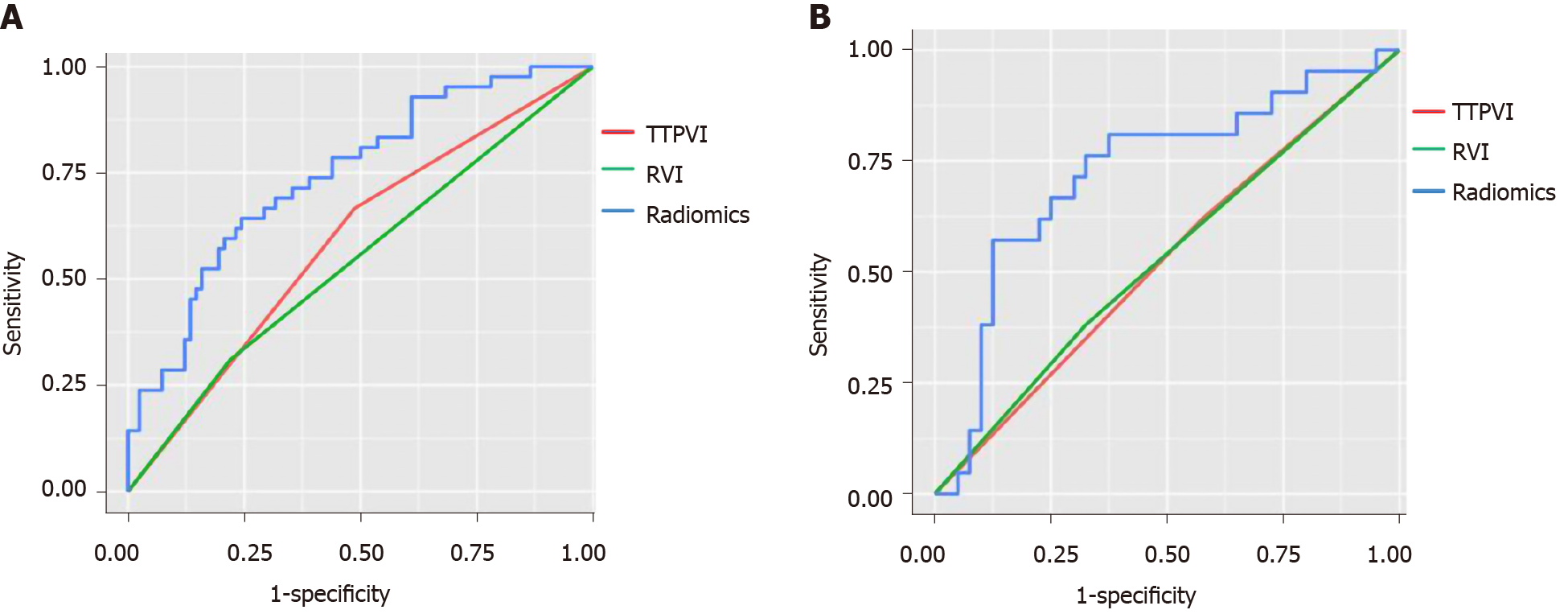
Figure 4 The two-trait predictor of venous invasion (green curve), radiogenomic invasion (red curve), and receiver operator characteristic of radiomics tag (blue curve) from the two groups.
A: Training group; B: Verification group. TTPVI: Two-trait predictor of venous invasion; RVI: Radiogenomic invasion.
- Citation: Liu P, Tan XZ, Zhang T, Gu QB, Mao XH, Li YC, He YQ. Prediction of microvascular invasion in solitary hepatocellular carcinoma ≤ 5 cm based on computed tomography radiomics. World J Gastroenterol 2021; 27(17): 2015-2024
- URL: https://www.wjgnet.com/1007-9327/full/v27/i17/2015.htm
- DOI: https://dx.doi.org/10.3748/wjg.v27.i17.2015


