Copyright
©The Author(s) 2021.
World J Gastroenterol. Apr 21, 2021; 27(15): 1578-1594
Published online Apr 21, 2021. doi: 10.3748/wjg.v27.i15.1578
Published online Apr 21, 2021. doi: 10.3748/wjg.v27.i15.1578
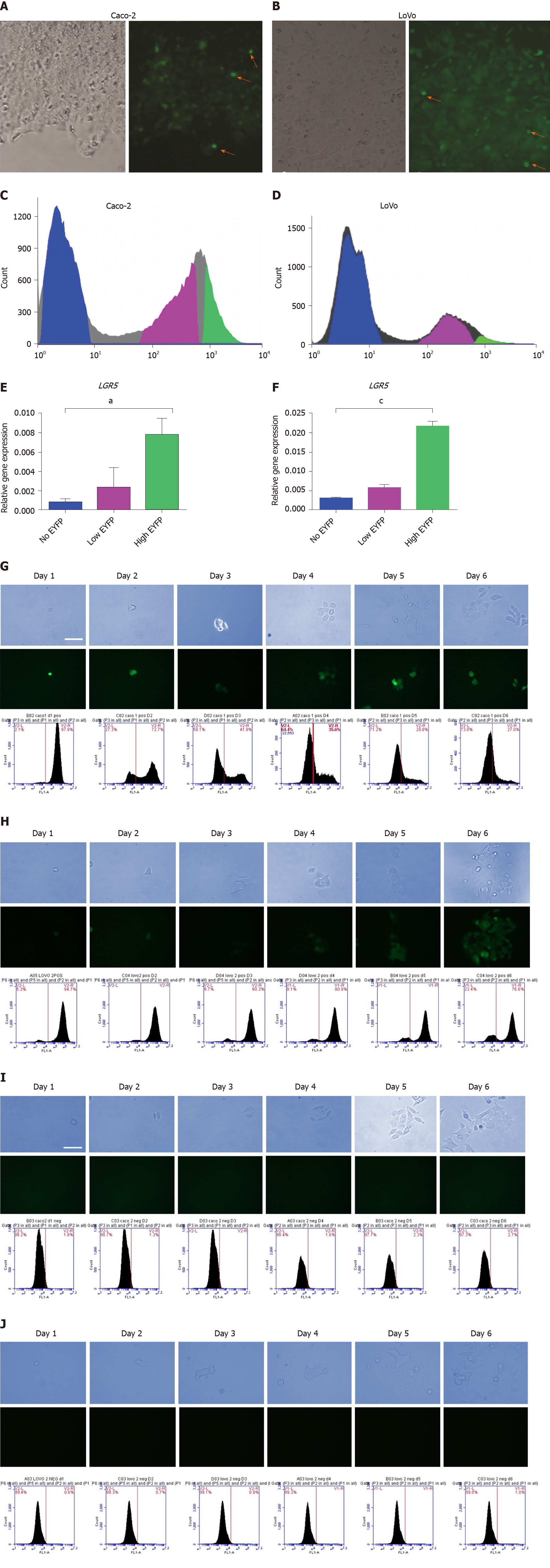
Figure 1 Proliferation and differentiation analysis of the stem cell compartment in stably transfected clones.
A and B: Phase contrast and fluorescence images show colonies of stably transfected Caco-2 (A) and LoVo (B) cells expressing the enhanced yellow fluorescent protein (EYFP) reporter (red arrows show small cells expressing high level of enhanced yellow fluorescent protein, EYFP); C and E: Fluorescence-activated cell sorting (FACS) sorting of leucine-rich repeat-containing G protein-coupled receptor 5 (LGR5)/EYFP expression in Caco-2 clone (C) and LoVo clones (E) into EYFPhigh, EYFPlow, and EYFPnegative populations; D and F: Quantitative real-time polymerase chain reaction analysis of LGR5 messenger ribonucleic acid expression in EYFPhigh, EYFPlow, and EYFPnegative cells relative to glyceraldehyde-3-phosphate dehydrogenase gene in Caco-2 clones (D) and LoVo clones (F); G: Phase contrast images show proliferation of EYFPhigh cells from LGR5/EYFP Caco-2 clone; fluorescence images show self-renewal of LGR5/EYFP stem cells and down-regulation of LGR5/EYFP in differentiated progeny in LGR5/EYFP Caco-2. Sequential FACS analysis shows EYFP expression in LGR5/EYFP Caco-2 clone over 6 d in culture; H: Phase contrast images show proliferation of EYFPnegative and cells from LGR5/EYFP Caco-2 clone; fluorescence images show self-renewal of LGR5 stem cells and down-regulation of LGR5 in differentiated progeny in LGR5/EYFP Caco-2. Sequential FACS analysis shows EYFP expression in LGR5/EYFP Caco-2 clone over 6 d in culture; I and J: Phase contrast images show proliferation of EYFPhigh (I) and EYFPnegative (J) cells from LGR5/EYFP LoVo clone; fluorescent pictures showing self-renewal of LGR5 stem cells and down-regulation of LGR5/EYFP in differentiated progeny in LGR5/EYFP LoVo; sequential FACS analysis of EYFP expression in LGR5/EYFP LoVo clone over 6 d in culture. LGR5: Leucine-rich repeat-containing G protein-coupled receptor 5; EYFP: Enhanced yellow fluorescent protein.
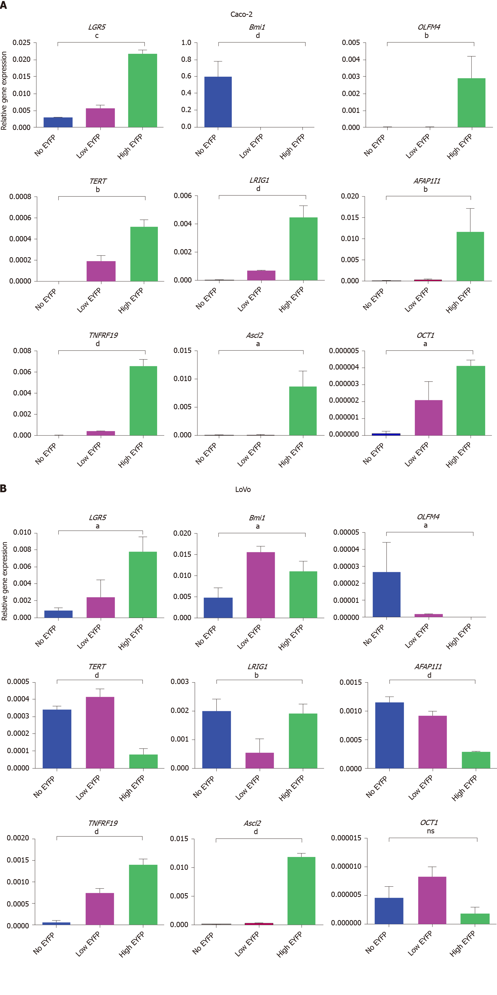
Figure 2 Quantitative polymerase chain reaction analysis.
A and B: Quantitative polymerase chain reaction analysis of intestinal stem cell markers and the cancer stem cell marker organic cation transporter 1 in leucine-rich repeat-containing G protein-coupled receptor 5/enhanced yellow fluorescent protein (LGR5/EYFP)high, LGR5/EYFPlow, and LGR5/EYFPnegative fractions of the Caco-2 cell line (A) and the LoVo cell line (B). The expression value of each gene was normalized to glyceraldehyde-3-phosphate dehydrogenase gene. Data were then analyzed by one-way analysis of variance. aP < 0.05, bP < 0.01, cP < 0.001, dP < 0.0001. LGR5: Leucine-rich repeat-containing G protein-coupled receptor 5; EYFP: Enhanced yellow fluorescent protein; Bmi1: B lymphoma Moloney murine leukemia virus insertion region 1 homolog; OCT1: organic cation transporter 1; TERT: Telomerase reverse transcriptase; ASCL2: Achaete-scute complex homolog 2.
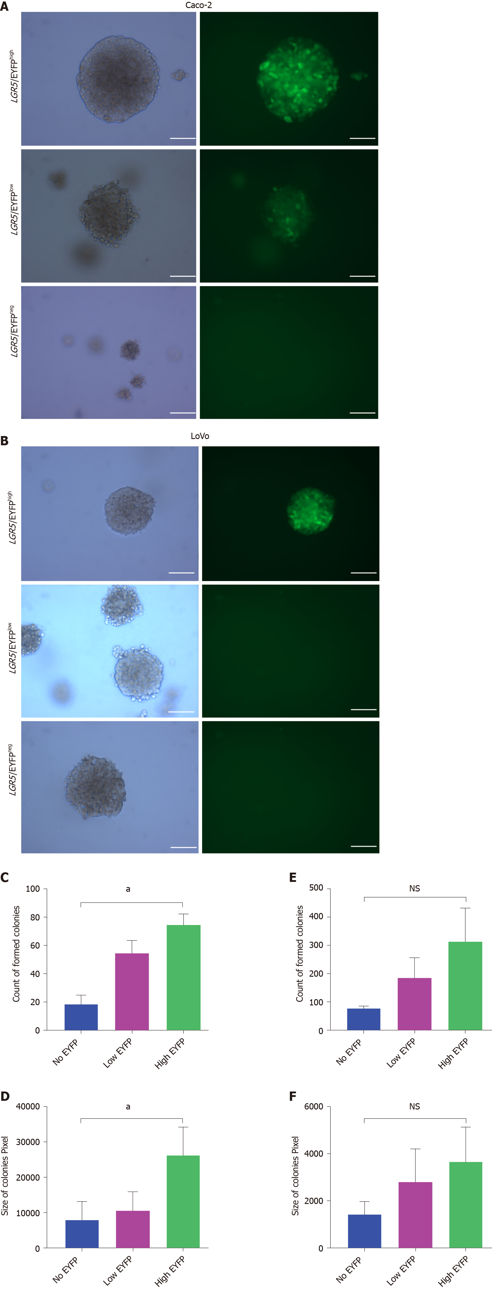
Figure 3 Invitro anchor independent assay of Caco-2 and LoVo colon cancer cell line.
Sorted cells were cultured for 2 wk in soft agar before analysis. A-F: Representative images of colonies formed by cells with different leucine-rich repeat-containing G protein-coupled receptor 5/enhanced yellow fluorescent protein levels in Caco-2 cell line (A) and in LoVo cell line (B). Scale bar, 50 µm; the (C) and (E) number, and (D) and (F) size of colonies formed by Caco-2 and LoVo clones respectively were assessed using crystal violet staining. Colonies larger than 0.25 mm in diameter were then counted. Columns and error bars represent means ± SD of two independent experiments using duplicate measurements in each experiment. aP < 0.05. LGR5: Leucine-rich repeat-containing G protein-coupled receptor 5; YFP: Yellow fluorescent protein; EYFP: Enhanced yellow fluorescent protein.
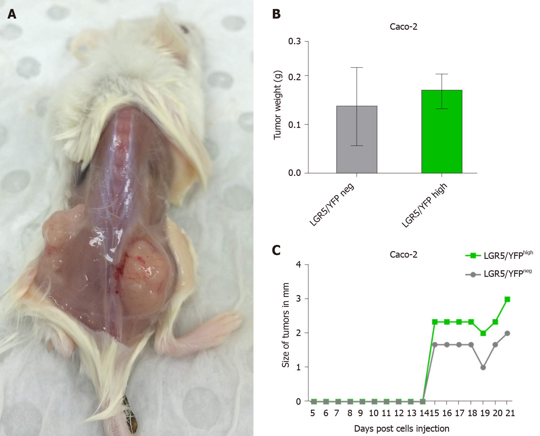
Figure 4 Stable leucine-rich repeat-containing G protein-coupled receptor 5/enhanced yellow fluorescent proteinhigh expressing Caco-2 cells line possesses stem cell-like properties in vivo.
Sorted leucine-rich repeat-containing G protein-coupled receptor 5/enhanced yellow fluorescent protein (LGR5/EYFP)negative and LGR5/EYFPhigh cells were injected subcutaneously in left and right flanks (respectively) of non-obese diabetic- severe combined immunodeficient mice and monitored for 3 wk. A: Representative images of tumors formed by LGR5/EYFPhigh (right flank) and LGR5/EYFPnegative (left flank) cells; B and C: Tumor weights (B) and size (C) resulting from LGR5/EYFPnegative and LGR5/EYFPhigh sorted cells. Columns and error bars represent means ± SD of two independent experiments using duplicate measurements for each experiment. neg: Negative; LGR5: Leucine-rich repeat-containing G protein-coupled receptor 5; YFP: Yellow fluorescent protein.
- Citation: Alharbi SA, Ovchinnikov DA, Wolvetang E. Leucine-rich repeat-containing G protein-coupled receptor 5 marks different cancer stem cell compartments in human Caco-2 and LoVo colon cancer lines. World J Gastroenterol 2021; 27(15): 1578-1594
- URL: https://www.wjgnet.com/1007-9327/full/v27/i15/1578.htm
- DOI: https://dx.doi.org/10.3748/wjg.v27.i15.1578


