Copyright
©The Author(s) 2021.
World J Gastroenterol. Jan 7, 2021; 27(1): 69-79
Published online Jan 7, 2021. doi: 10.3748/wjg.v27.i1.69
Published online Jan 7, 2021. doi: 10.3748/wjg.v27.i1.69
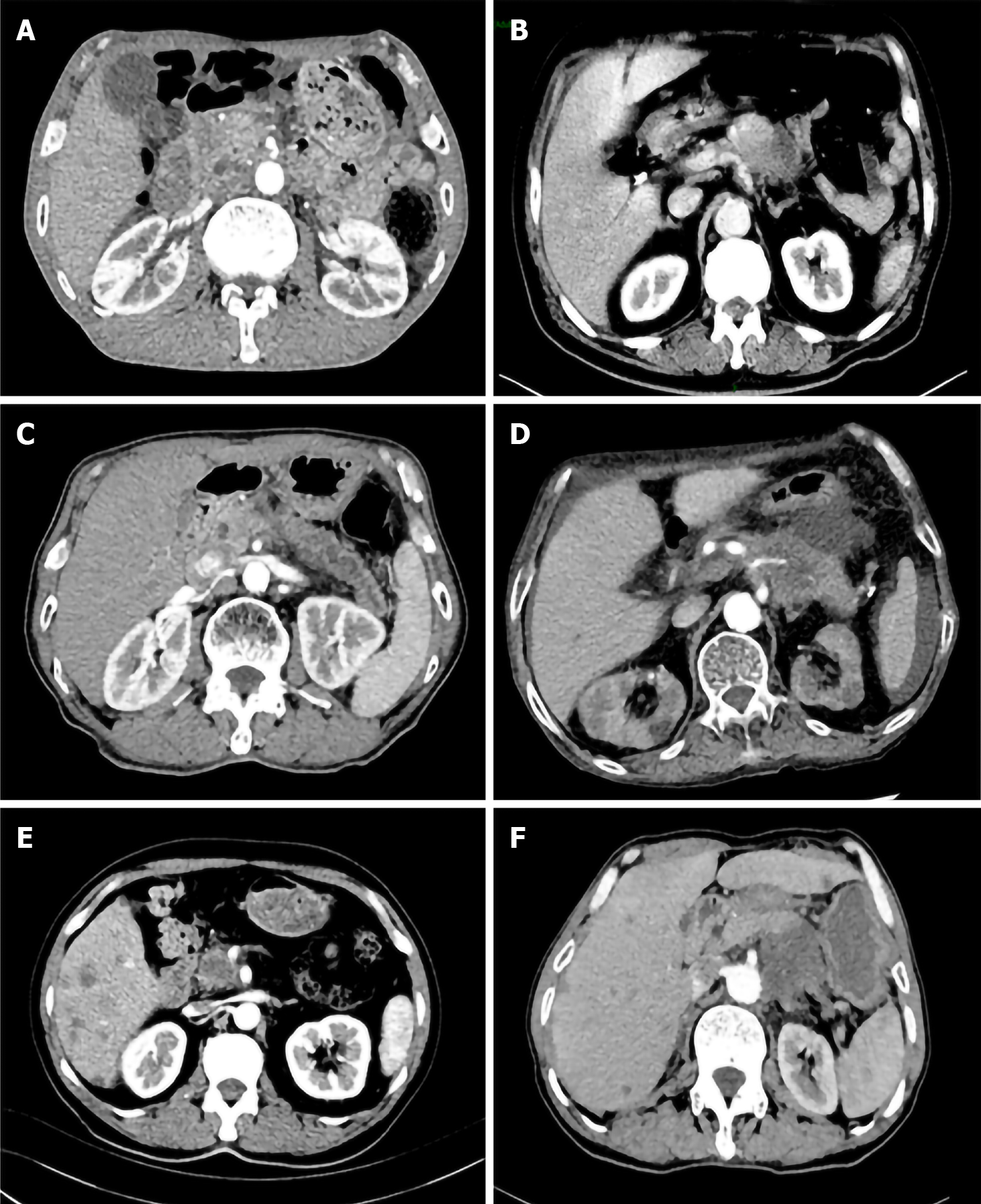
Figure 1 Varied pancreatic lesions are shown by contrast-enhanced computed tomography image.
A: The lesion was located in head/neck of pancreas; B: The lesion was located in body/tail of pancreas; C: Pancreatic head lesion was associated with celiac trunk and celiac plexus invasion; D: The image showed a pancreatic body/tail invading the celiac plexus; E: Pancreatic head/neck lesion was accompanied with hepatic metastasis; F: Pancreatic body/tail lesion was accompanied with hepatic metastasis.
- Citation: Han CQ, Tang XL, Zhang Q, Nie C, Liu J, Ding Z. Predictors of pain response after endoscopic ultrasound-guided celiac plexus neurolysis for abdominal pain caused by pancreatic malignancy. World J Gastroenterol 2021; 27(1): 69-79
- URL: https://www.wjgnet.com/1007-9327/full/v27/i1/69.htm
- DOI: https://dx.doi.org/10.3748/wjg.v27.i1.69


