Copyright
©The Author(s) 2021.
World J Gastroenterol. Jan 7, 2021; 27(1): 129-142
Published online Jan 7, 2021. doi: 10.3748/wjg.v27.i1.129
Published online Jan 7, 2021. doi: 10.3748/wjg.v27.i1.129
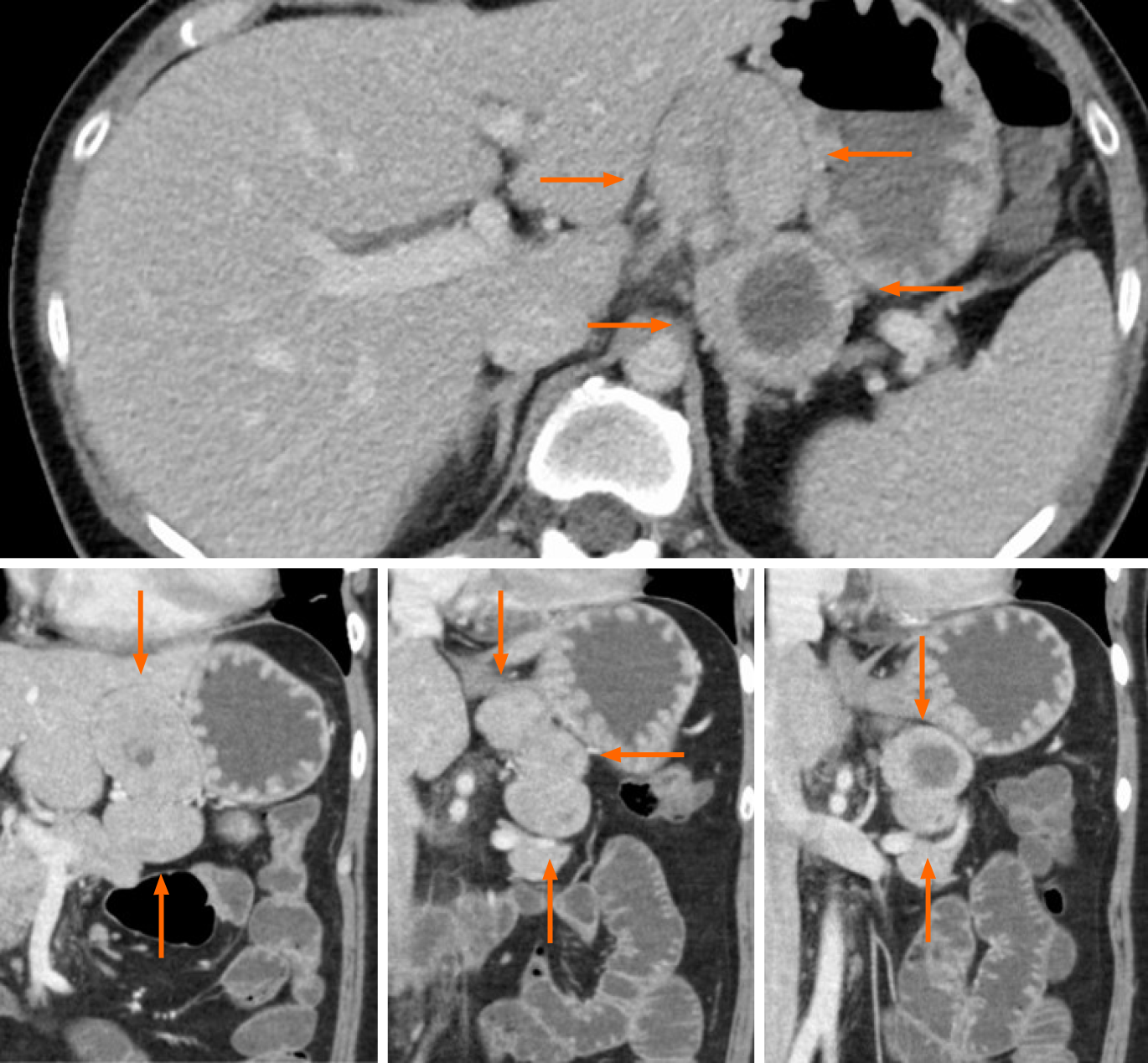
Figure 1 Axial (top) and coronal (bottom) views of computed tomography with contrast at admission revealed an incidental 8 cm mass in the lesser omentum (orange arrows).
The mass appeared to result from the fusion of 3 similar tumors, of which 1 contained a non-enhancing, low-density area suggestive of necrosis or hematoma.
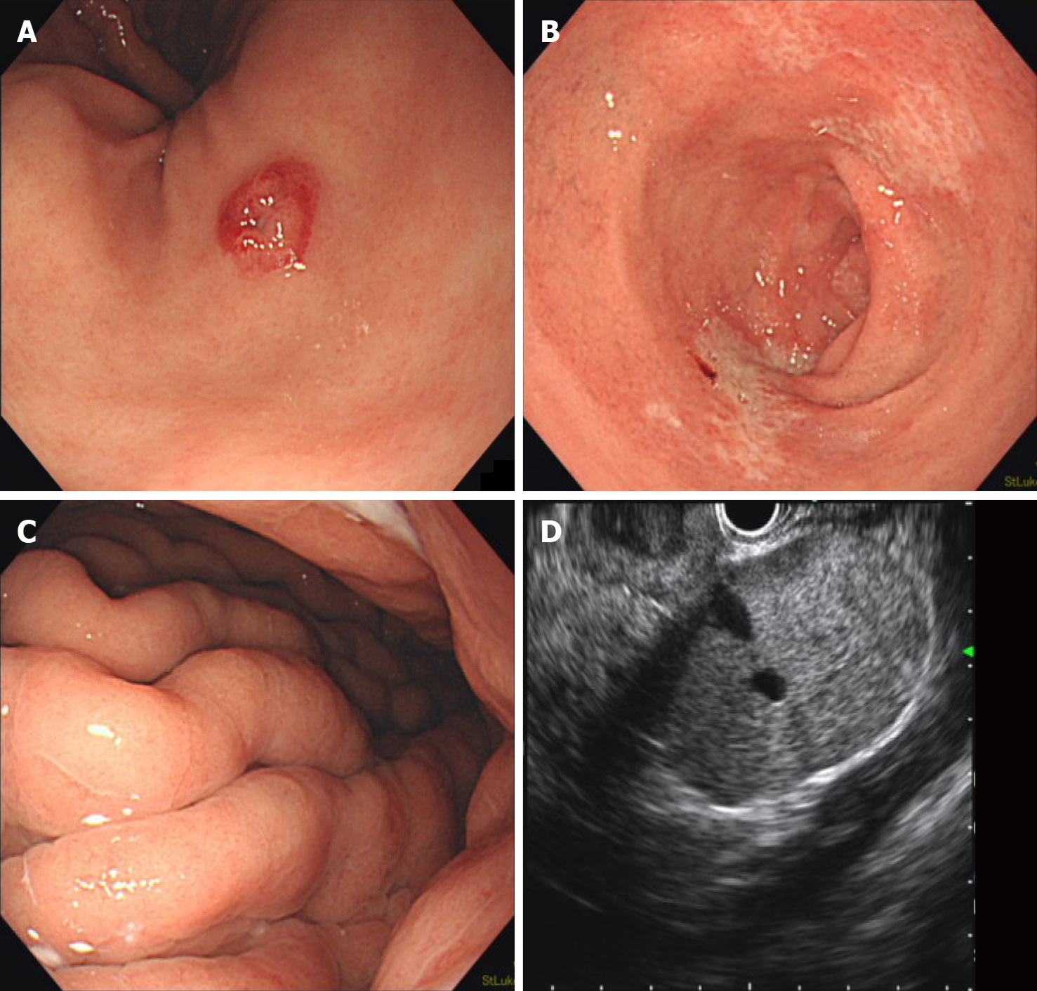
Figure 2 Esophagogastroduodenoscopy incidentally revealed a red, 7 mm submucosal tumor with a central depression (A); prominent gastric folds (B) and shallow duodenal ulcers (C) were also noted; endoscopic ultrasound revealed 3 clearly delineated, hyperechoic masses with uniform texture, of which 1 had a hypoechoic center with clear borders (D).
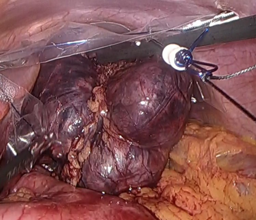
Figure 3 Laparoscopic surgery revealed an 83 mm × 80 mm × 37 mm mass in the lesser omentum which appeared to be formed from the fusion of 3 spherical tumors.
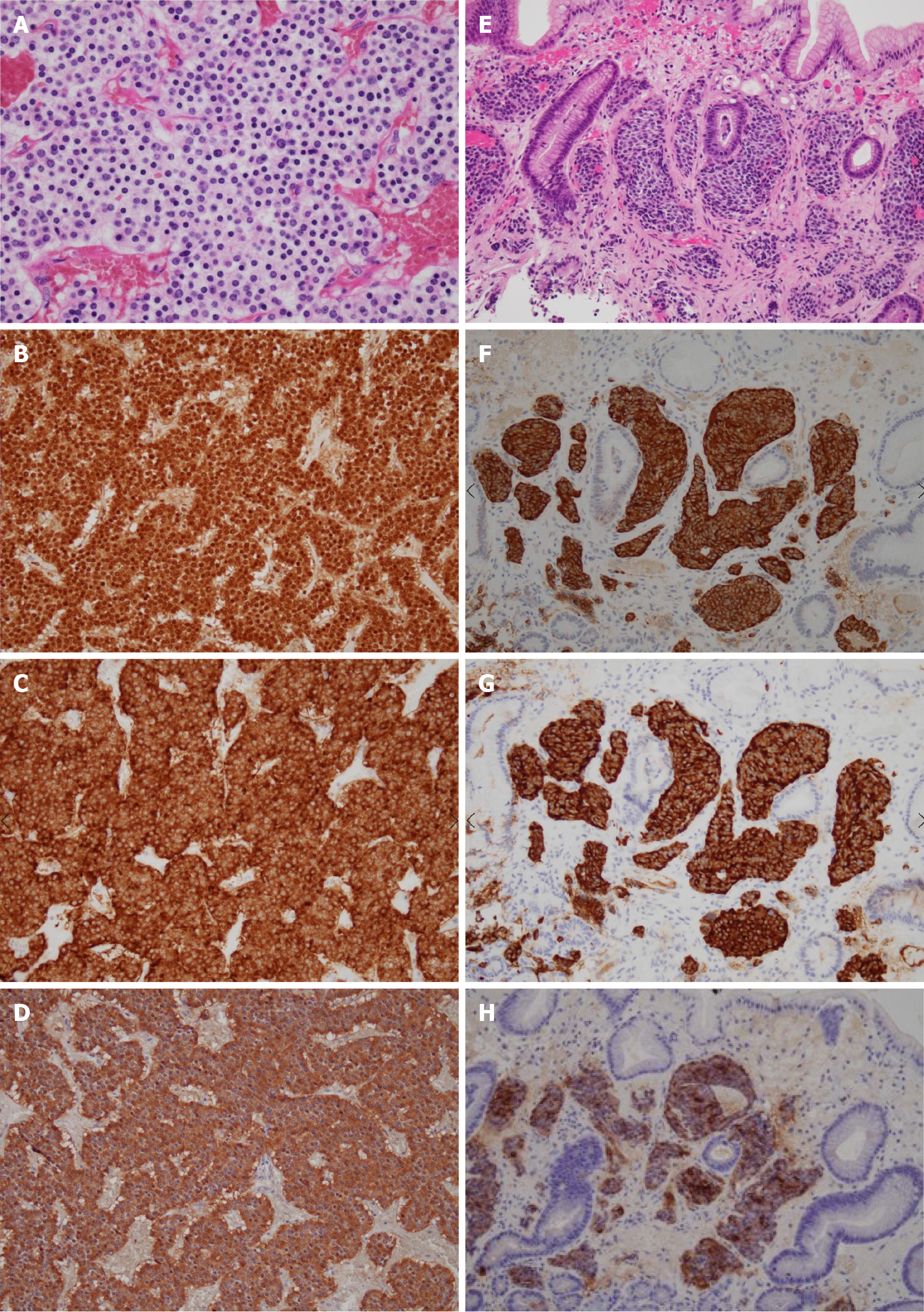
Figure 4 Pathology of the surgical specimen (A-D) and gastric biopsy (E-H).
Nests of tumor cells characterized by small ovoid nuclei and mildly eosinophilic cytoplasms with intervening dilated capillary networks were observed in the omental lesion (A). The tumor was positive for chromogranin A (B), synaptophysin (C), and gastrin (D) stains. Biopsy of the gastric lesion showed similar cells in the mucosal layer (E) which were also positive for chromogranin A (F), synaptophysin (G), and gastrin (H) stains.
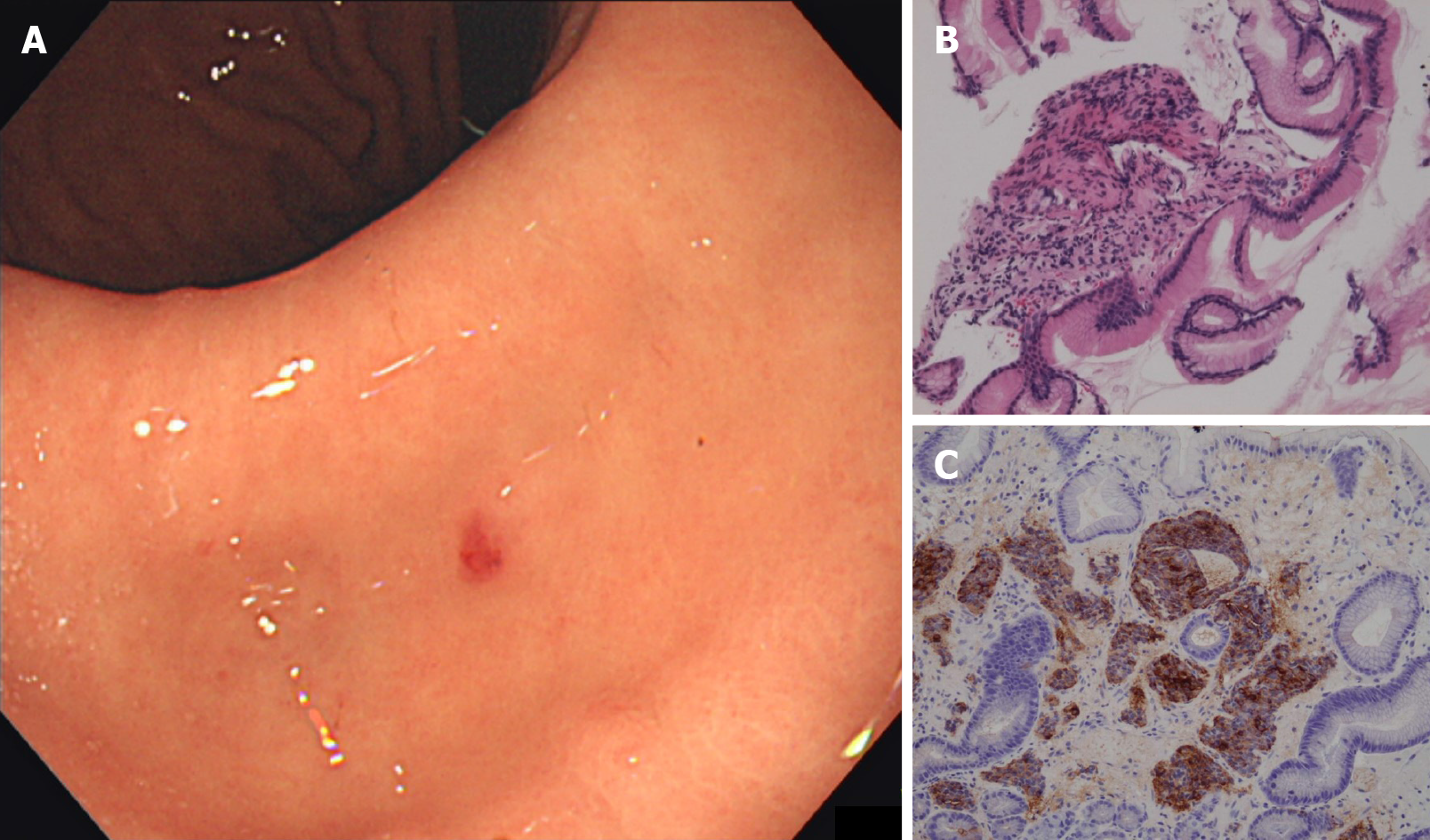
Figure 5 The gastric neuroendocrine neoplasm was barely identifiable on follow-up esophagogastroduodenoscopy, reducing to a red dot with no visible elevation (A); biopsy of the lesion was negative for tumor, with only regenerative and fibrous changes (B); synaptophysin (C) stain was also negative.
- Citation: Okamoto T, Yoshimoto T, Ohike N, Fujikawa A, Kanie T, Fukuda K. Spontaneous regression of gastric gastrinoma after resection of metastases to the lesser omentum: A case report and review of literature. World J Gastroenterol 2021; 27(1): 129-142
- URL: https://www.wjgnet.com/1007-9327/full/v27/i1/129.htm
- DOI: https://dx.doi.org/10.3748/wjg.v27.i1.129


