Copyright
©The Author(s) 2020.
World J Gastroenterol. Feb 28, 2020; 26(8): 877-882
Published online Feb 28, 2020. doi: 10.3748/wjg.v26.i8.877
Published online Feb 28, 2020. doi: 10.3748/wjg.v26.i8.877
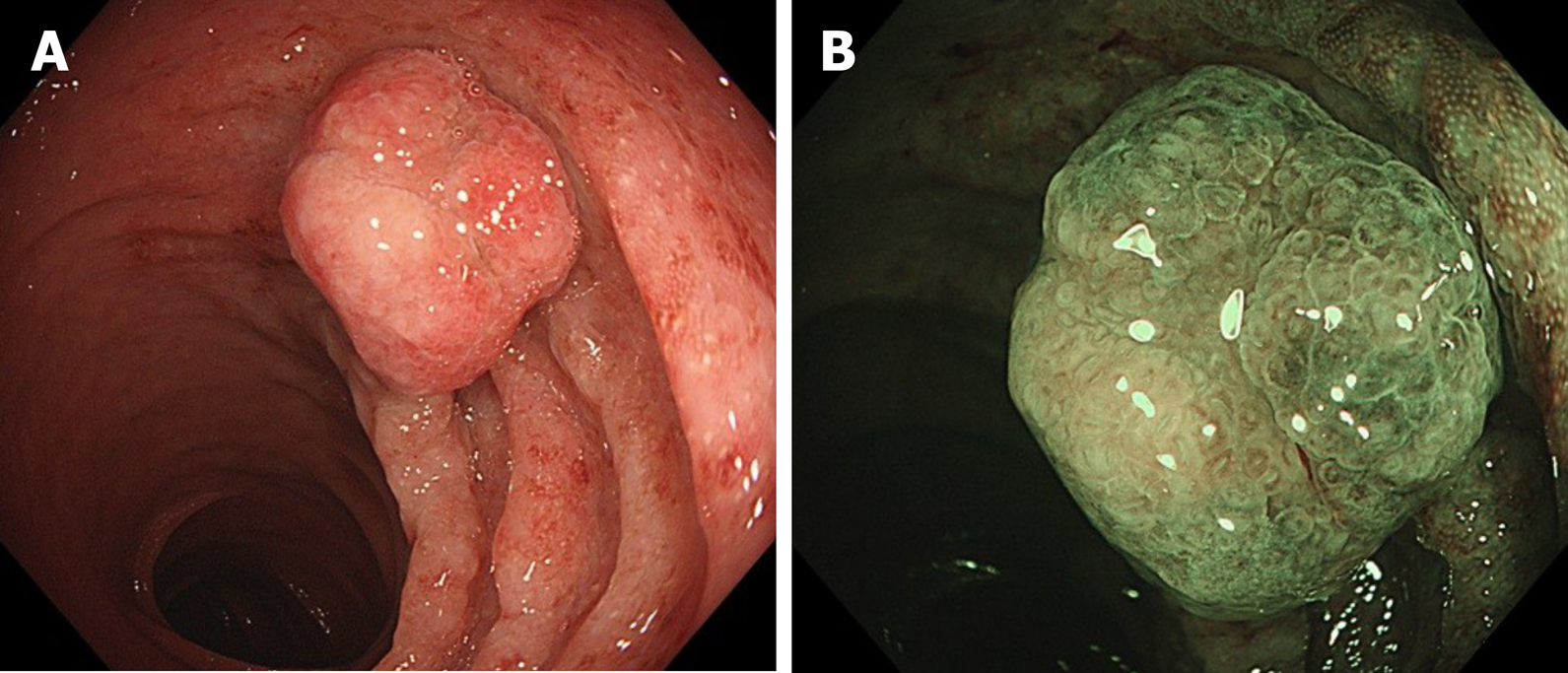
Figure 1 Endoscopic illustrations (CF-HQ 290, Olympus).
A: White light endoscopy; B: Narrow banded imaging endoscopy.
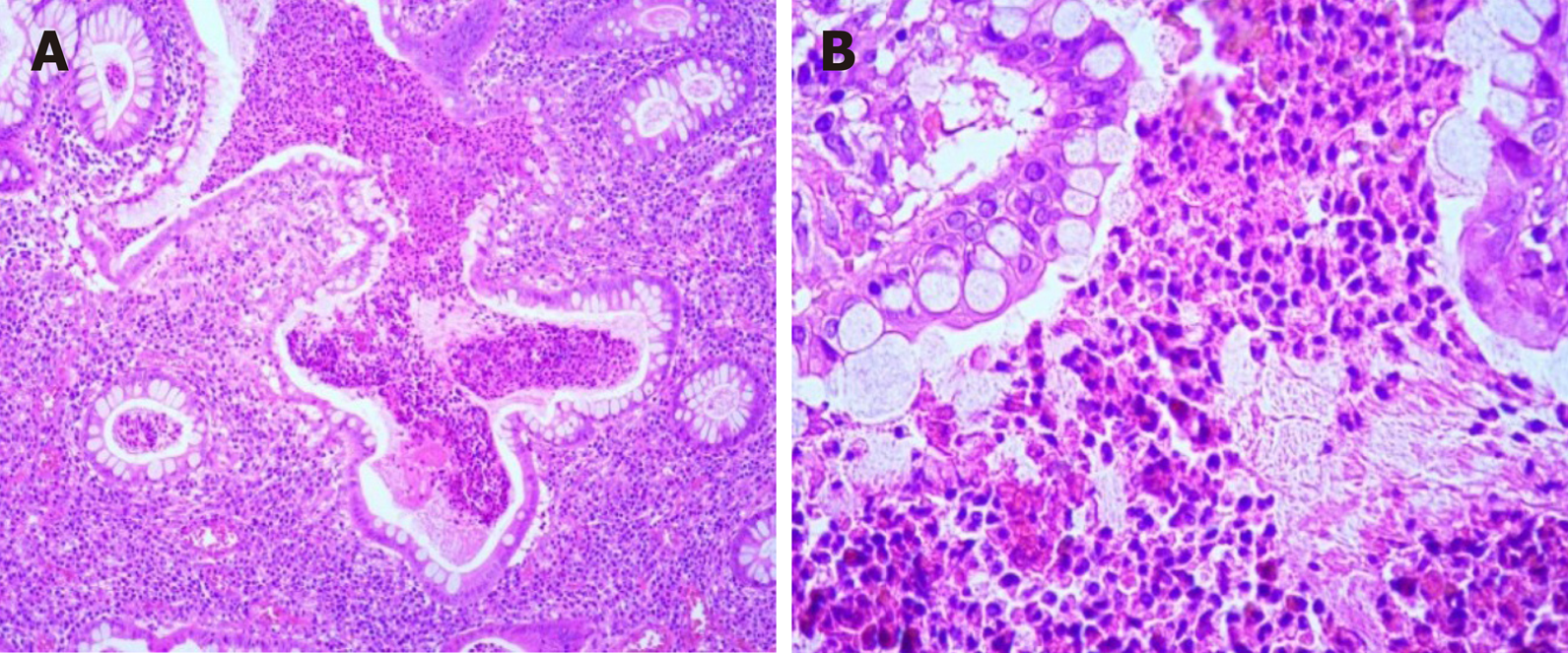
Figure 2 Hematoxylin and eosin staining of the juvenile polyp showed dilated hypersecreting glands.
A: × 10; B: × 40.
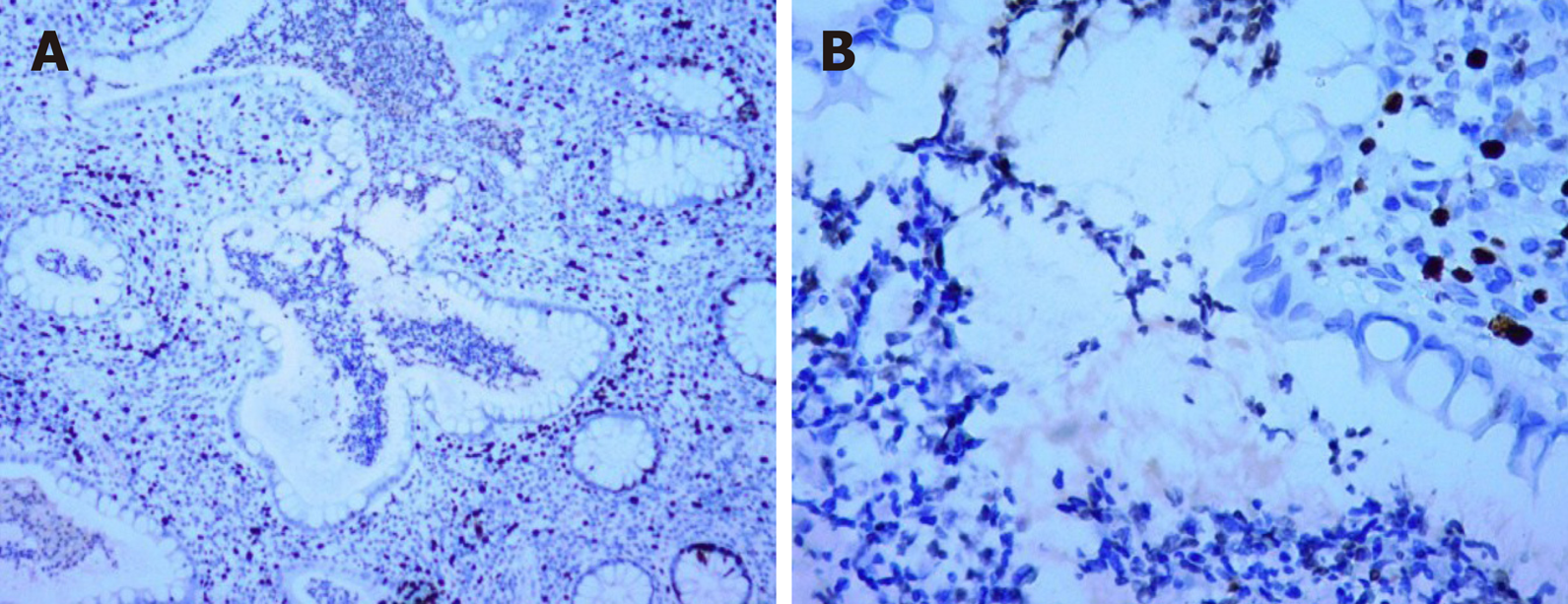
Figure 3 Immunohistochemical staining for p53 in the juvenile polyp showed wild-type expression and no overexpression.
A: × 10; B: × 40.
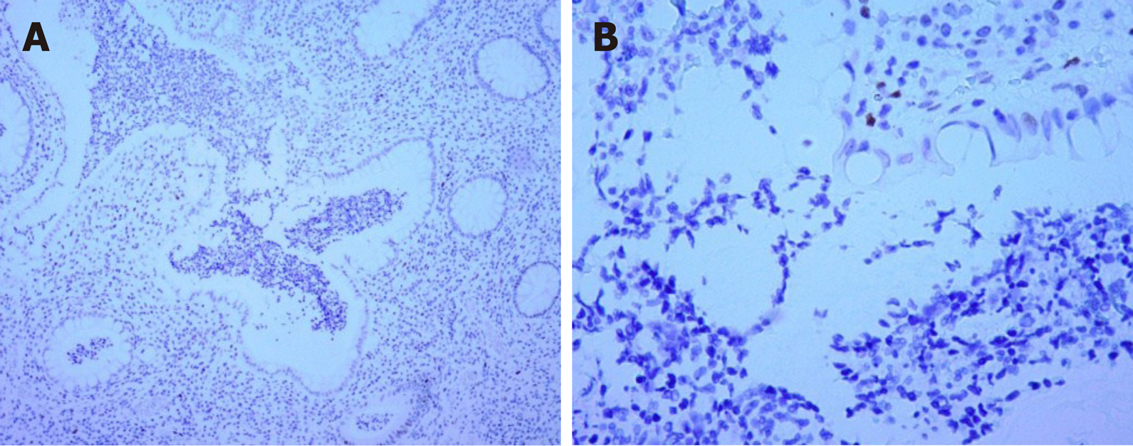
Figure 4 Immunohistochemical staining for Ki-67 showed that the Ki-67 labeling index was 3%.
A: × 10; B: × 40.
- Citation: Chen YW, Tu JF, Shen WJ, Chen WY, Dong J. Diagnosis and management of a solitary colorectal juvenile polyp in an adult during follow-up for ulcerative colitis: A case report. World J Gastroenterol 2020; 26(8): 877-882
- URL: https://www.wjgnet.com/1007-9327/full/v26/i8/877.htm
- DOI: https://dx.doi.org/10.3748/wjg.v26.i8.877


