Copyright
©The Author(s) 2020.
World J Gastroenterol. Dec 28, 2020; 26(48): 7603-7618
Published online Dec 28, 2020. doi: 10.3748/wjg.v26.i48.7603
Published online Dec 28, 2020. doi: 10.3748/wjg.v26.i48.7603
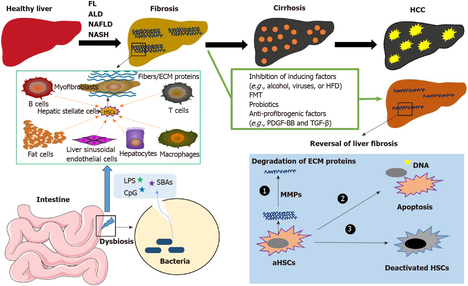
Figure 1 The development of liver diseases.
Without effective treatment or preventive strategies, fatty liver disease, alcohol-induced liver disease, nonalcoholic fatty liver disease, and nonalcoholic steatohepatitis can result in liver fibrosis, cirrhosis, and hepatocellular carcinoma. Gut microbiota-derived molecules, including lipopolysaccharide, CpG, and secondary bile acids, initially activate liver resident cells to produce cytokines and chemokines. Quiescent hepatic stellate cells (HSCs) can be activated and transformed into myofibroblasts (MFBs), which is mediated by chemokines and cytokines released by liver-infiltrating macrophages, leukocytes, and other cell types, including fat cells and damaged hepatocytes. MFBs are the predominant source of collagen-producing cells and other extracellular matrix proteins (ECM). With effective treatment, such as fecal microbiota transplant, probiotics, and anti-profibrogenic factors, fibrosis is reversible. The treatments that induce apoptosis (or deactivation) of activated HSCs or MFBs and degrade the ECM proteins can reduce the stiffness of the liver, reverse liver fibrosis, and inhibit the progression of liver disease. FL: Fatty liver; ALD: Alcohol-induced liver disease; NAFLD: Nonalcoholic fatty liver disease; NASH: Nonalcoholic steatohepatitis; HCC: Hepatocellular carcinoma; ECM: Extracellular matrix proteins; HFD: High-fat diet; FMT: Fecal microbiota transplant; LPS: Lipopolysaccharide; SBAs: Secondary bile acids; HSC: Hepatic stellate cells.
- Citation: Qi X, Yang M, Stenberg J, Dey R, Fogwe L, Alam MS, Kimchi ET, Staveley-O'Carroll KF, Li G. Gut microbiota mediated molecular events and therapy in liver diseases. World J Gastroenterol 2020; 26(48): 7603-7618
- URL: https://www.wjgnet.com/1007-9327/full/v26/i48/7603.htm
- DOI: https://dx.doi.org/10.3748/wjg.v26.i48.7603


