Copyright
©The Author(s) 2020.
World J Gastroenterol. Dec 7, 2020; 26(45): 7104-7117
Published online Dec 7, 2020. doi: 10.3748/wjg.v26.i45.7104
Published online Dec 7, 2020. doi: 10.3748/wjg.v26.i45.7104
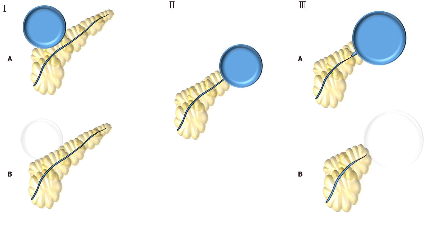
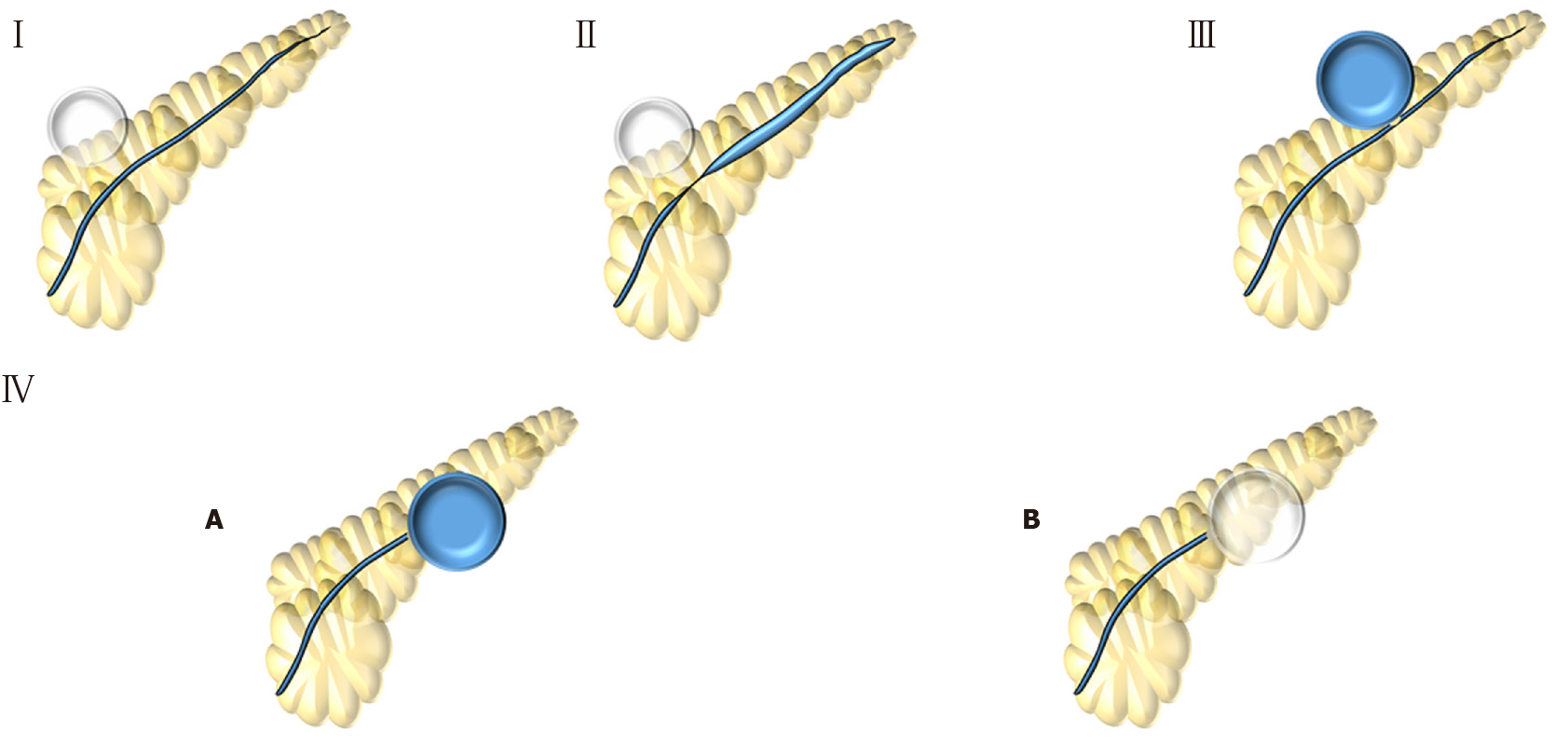
Figure 1 Nordback et al[7] (1988) classification.
Type I: Normal main pancreatic duct (MPD) contrasting (type IA) or not (type IB) the collection; Type II: MPD opens to the collection; Type III: MPD with stenosis contrasting (type IIIA) or not (type IIIB) the collection.
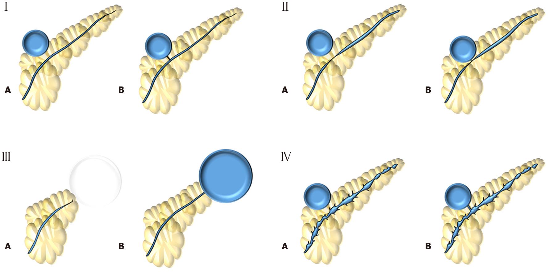
Figure 2 Nealon et al[37] (2009) classification.
Type I: Normal main pancreatic duct (MPD); Type II: MPD stricture; Type III: MPD occlusion; Type IV: Chronic pancreatitis. All types are subdivided according if they have communication (subtype A) or not (subtype B) with the collection.
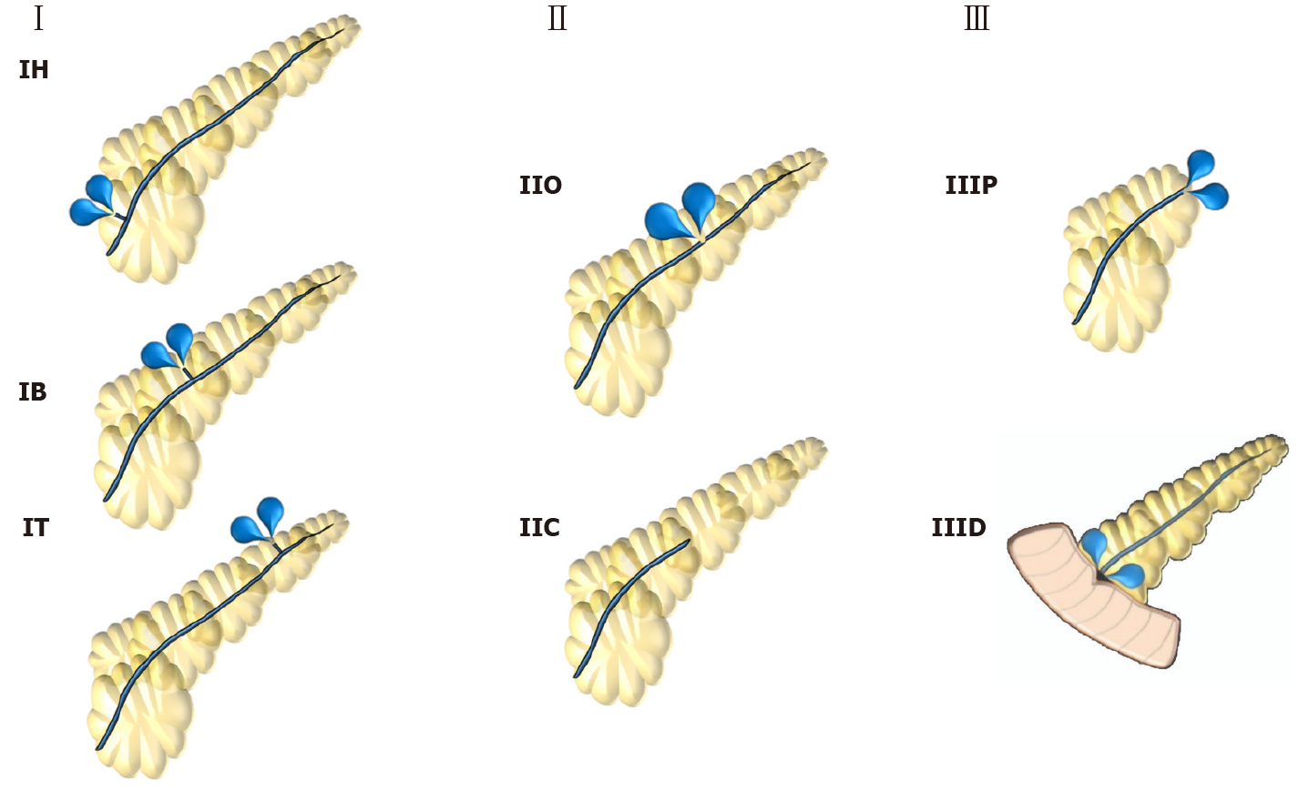
Figure 3 Mutignani et al[35] (2017) classification.
Type I: Leakages from small side brunches in the pancreatic head (IH), body (IB) or tail (IT); Type II: Leak in the main pancreatic duct that may have an open (IIO) or close (IIC) proximal stump; Type III: Leaks after pancreatectomy that may be after proximal pancreas (IIIP) or distal pancreas (IIID) resection.
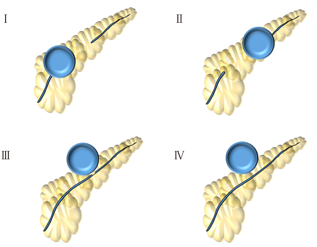
Figure 4 Dhir et al[23] (2018) classification.
Type I: Disconnection in the neck/body region, with a ductal leak at the proximal end; Type II: Disconnected duct with a Walled-off Necrosis distal to the disconnection – not possible to ascertain ductal communication with collection; Type III: Ductal leak without disconnection; Type IV: Shows a noncommunicating Walled-off Necrosis, with no disconnection.
Figure 5 Lera-Proença (2020) new proposed classification.
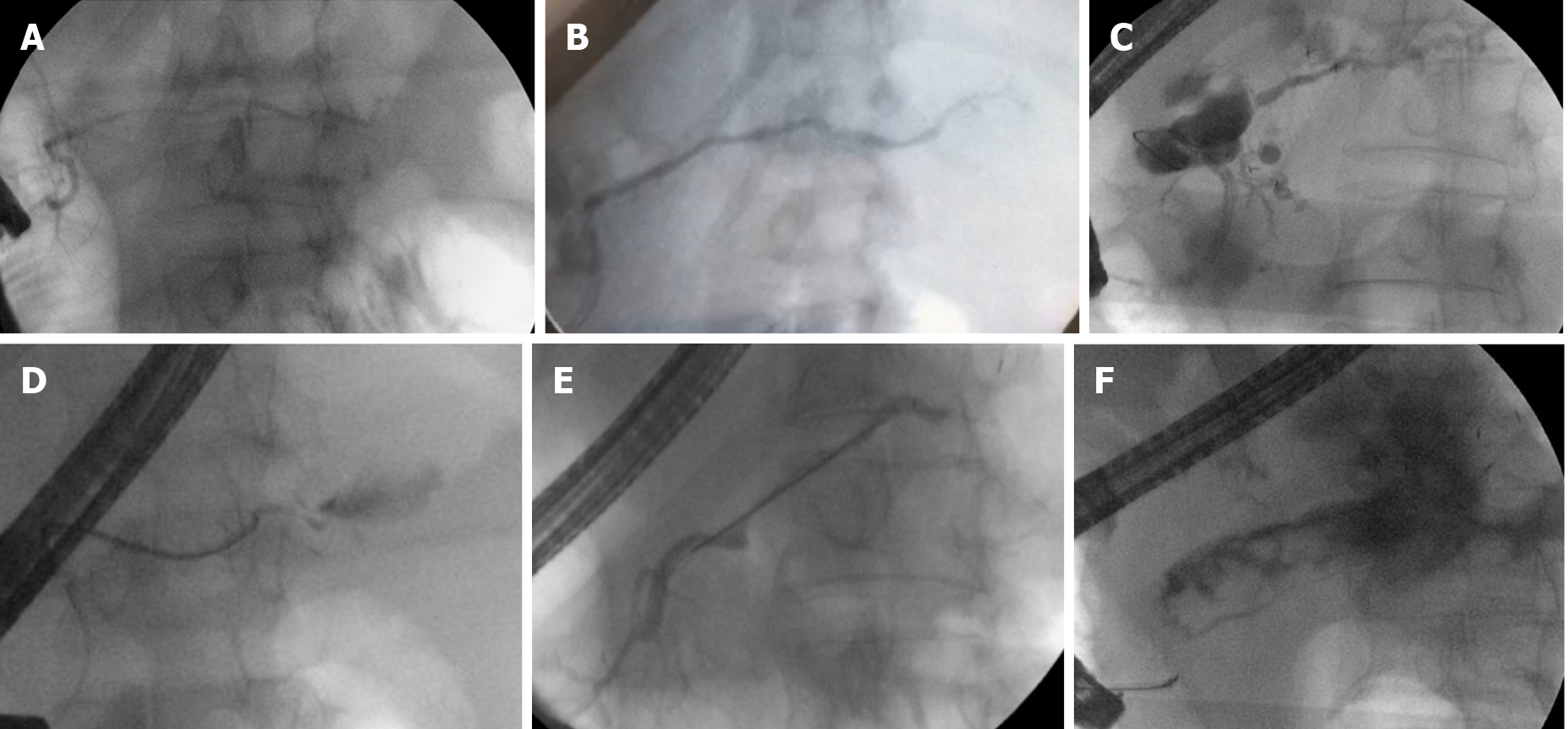
Type I: Normal main pancreatic duct; Type II: Stricture; Type III: Partial disruption – main pancreatic duct contrasts beyond disruption; Type IV: Complete disruption - main pancreatic duct does not contrast beyond disruption. IV-A: with contrast extravasation or IV-B: without contrast extravasation and cut-off.
Figure 6 Endoscopic pancreatography classified by Lera-Proença classification.
Endoscopic pancreatography findings, A: Normal pancreatography (type I); B: Stricture (type II); C: Partial disruption (type III); D: Complete disruption with contrast extravasation (type IV-A); E: Complete disruption without contrast extravasation and cut-off (Type IV-B); and F: Stricture and complete disruption with contrast extravasation (Type II + IV-A).
- Citation: Proença IM, dos Santos MEL, de Moura DTH, Ribeiro IB, Matuguma SE, Cheng S, McCarty TR, do Monte Junior ES, Sakai P, de Moura EGH. Role of pancreatography in the endoscopic management of encapsulated pancreatic collections – review and new proposed classification. World J Gastroenterol 2020; 26(45): 7104-7117
- URL: https://www.wjgnet.com/1007-9327/full/v26/i45/7104.htm
- DOI: https://dx.doi.org/10.3748/wjg.v26.i45.7104


