Copyright
©The Author(s) 2020.
World J Gastroenterol. Nov 14, 2020; 26(42): 6658-6668
Published online Nov 14, 2020. doi: 10.3748/wjg.v26.i42.6658
Published online Nov 14, 2020. doi: 10.3748/wjg.v26.i42.6658
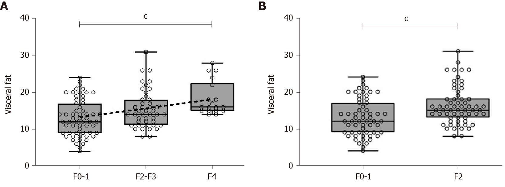
Figure 1 Visceral fat measurement by bioimpedanciometry, according to histological fibrosis stage.
A: Visceral fat measurements increased along with fibrosis stage assessed by histological analysis (F0-1, 12; F2-3, 14; F4, 16; Kruskal-Wallis cP < 0.001). A line can be fit by linear regression, showing linear association (r2 = 0.11, cP < 0.001); B: Visceral fat measurements were greater for those patients with significant fibrosis (16.3 vs 13.1, cP < 0.001).
Figure 2 Area under the receiver operating characteristic curve.
A: Receiver operating characteristic (ROC) curve for non-invasive diagnosis of significant liver fibrosis by a model including age and visceral fat; B: Comparison of the areas under ROC curves for a model using age and visceral fat versus liver elastography measurement, to predict significant liver fibrosis. Circles denote our model, triangles indicate non-alcoholic fatty liver disease fibrosis score and crosses denote liver elastography.
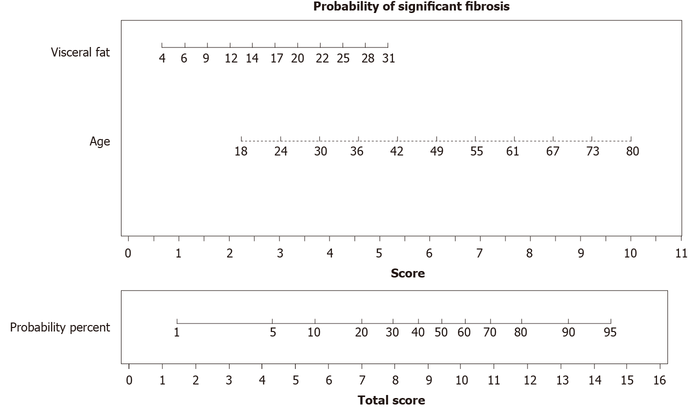
Figure 3 Nomogram for assessing the probability of significant liver fibrosis in a clinically useful manner.
With the variables resulting from the multivariate regression model, we built an easy-to-use visual tool. In an individual patient, visceral fat levels and age correspond to a score. Combining these scores gives a total score that can be converted to a probability of that patient having significant fibrosis in liver biopsy. For example, a patient with a visceral fat level of 12 (score 2) and with 55 years old (score 7) would have a total score of 9 and a corresponding probability of histological significant fibrosis of 43%.
- Citation: Hernández-Conde M, Llop E, Fernández Carrillo C, Tormo B, Abad J, Rodriguez L, Perelló C, López Gomez M, Martínez-Porras JL, Fernández Puga N, Trapero-Marugan M, Fraga E, Ferre Aracil C, Calleja Panero JL. Estimation of visceral fat is useful for the diagnosis of significant fibrosis in patients with non-alcoholic fatty liver disease. World J Gastroenterol 2020; 26(42): 6658-6668
- URL: https://www.wjgnet.com/1007-9327/full/v26/i42/6658.htm
- DOI: https://dx.doi.org/10.3748/wjg.v26.i42.6658


