Copyright
©The Author(s) 2020.
World J Gastroenterol. Oct 21, 2020; 26(39): 5997-6014
Published online Oct 21, 2020. doi: 10.3748/wjg.v26.i39.5997
Published online Oct 21, 2020. doi: 10.3748/wjg.v26.i39.5997
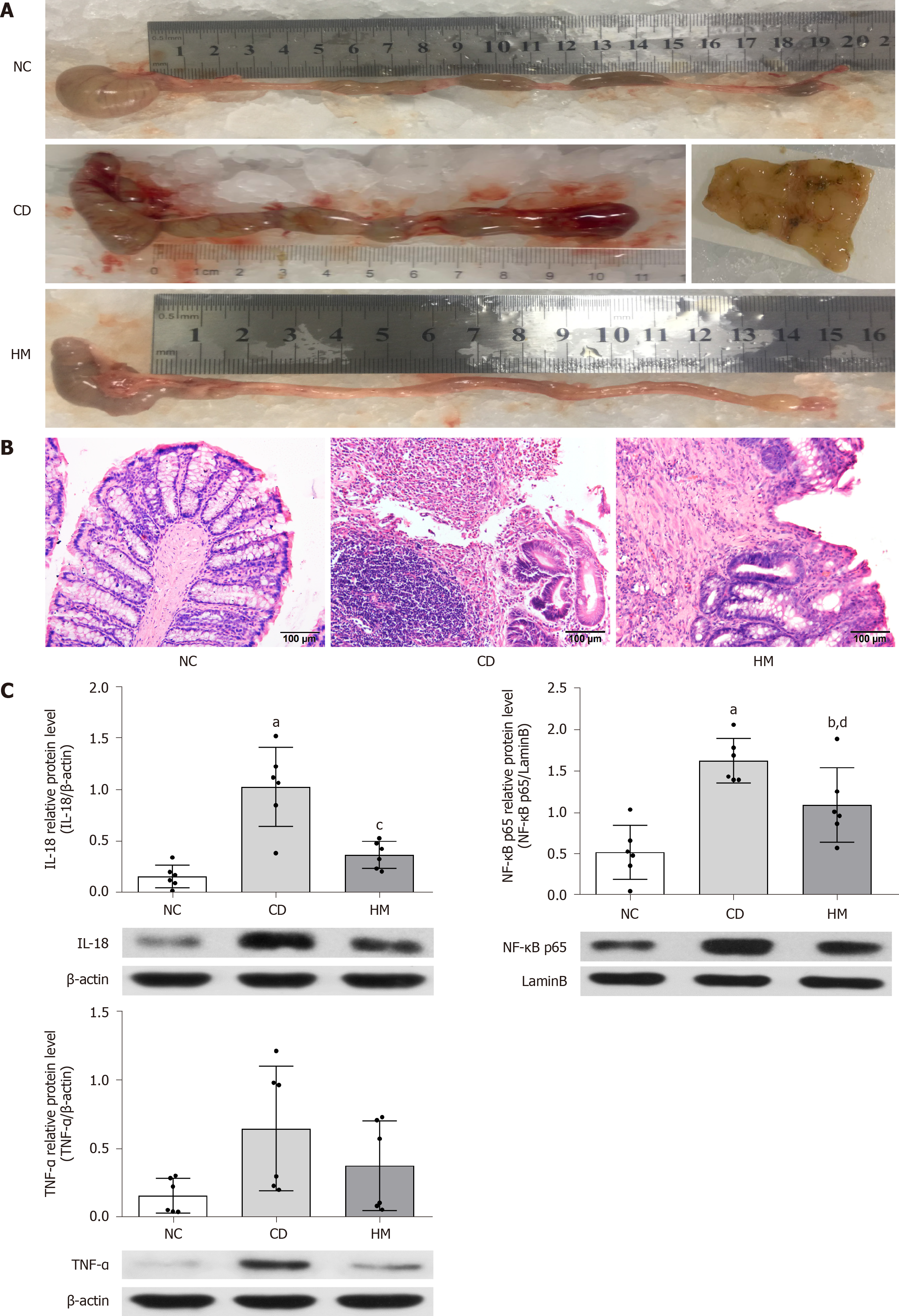
Figure 1 Herbal cake-partitioned moxibustion at the Qihai (CV6) and bilateral Tianshu (ST25) acupoints ameliorates colon damage and inflammation in Crohn’s disease rats.
A: Megacolon and cobblestone-like changes; B: Histopathological observations of colon tissue by hematoxylin and eosin staining. Scale bar: 100 μm; C: Expression of the colonic inflammatory factors interleukin-18 (IL-18), nuclear factor κB/p65 (NF-κB p65), and tumor necrosis factor-α (TNF-α) proteins evaluated by Western blot. Six independent experiments were analyzed, and the data are presented as the mean ± SD. aP < 0.01, bP < 0.05 vs NC group; cP < 0.01, dP < 0.05 vs CD group. NC: Normal control; CD: Crohn’s disease; HM: Herbal cake-partitioned moxibustion; IL-18: Interleukin 18; NF-κB p65: Nuclear factor κB p65; TNF-α: Tumor necrosis factor-α.
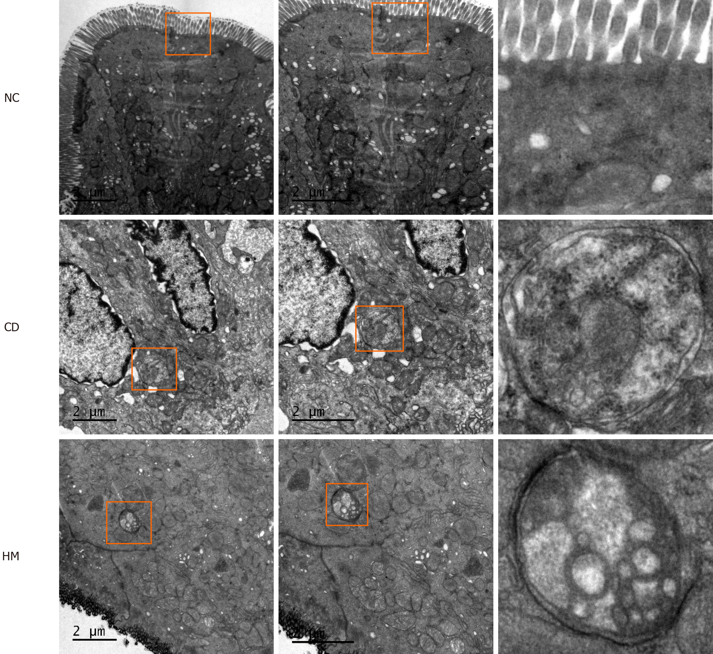
Figure 2 Ultrastructure of colonic epithelial tissue and autophagic vesicles in each group of rats.
Scale bar: 2 μm. NC: Normal control; CD: Crohn’s disease; HM: Herbal cake-partitioned moxibustion.
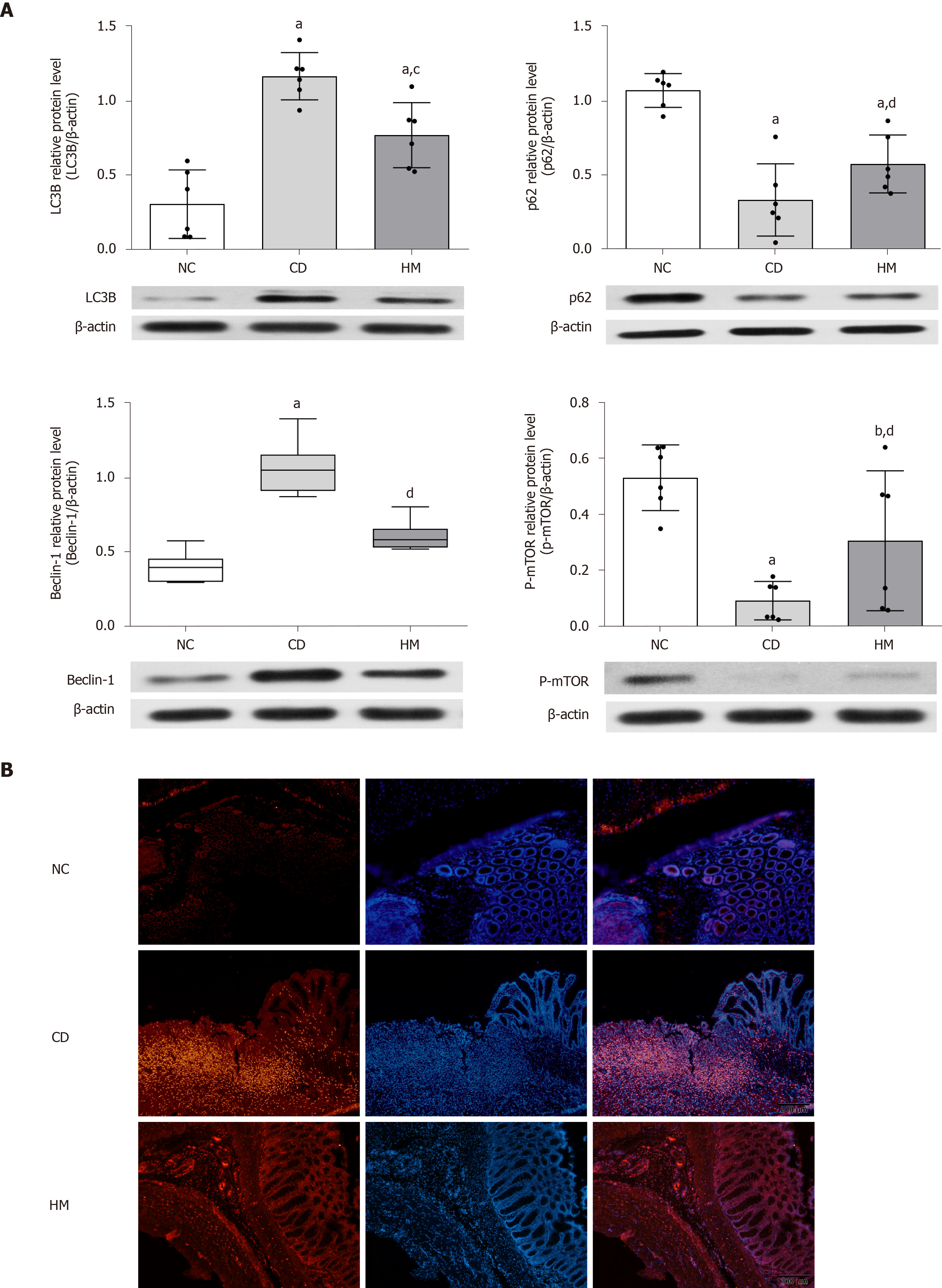
Figure 3 Herbal cake-partitioned moxibustion at the Qihai (CV6) and bilateral Tianshu (ST25) acupoints regulates the expression of autophagy proteins in the colon tissues of Crohn’s disease rats.
A: Expression changes of the microtubule-associated protein 1 light chain 3 beta, sequestosome 1, coiled-coil myosin-like BCL2-interacting protein, and phospho-mammalian target of rapamycin proteins evaluated by Western blot. Six independent experiments were analyzed, and the data are presented as the mean ± SD or medians (P25, P75). aP < 0.01, bP < 0.05 vs NC group; cP < 0.01, dP < 0.05 vs CD group; B: Immunofluorescence images for LC3B (Red) in each group. Scale bar: 200 μm. NC: Normal control; CD: Crohn’s disease; HM: Herbal cake-partitioned moxibustion; LC3B: Microtubule-associated protein 1 light chain 3 beta; p62: Sequestosome 1; Beclin-1: Coiled-coil myosin-like BCL2-interacting protein; p-mTOR: Phospho-mammalian target of rapamycin.
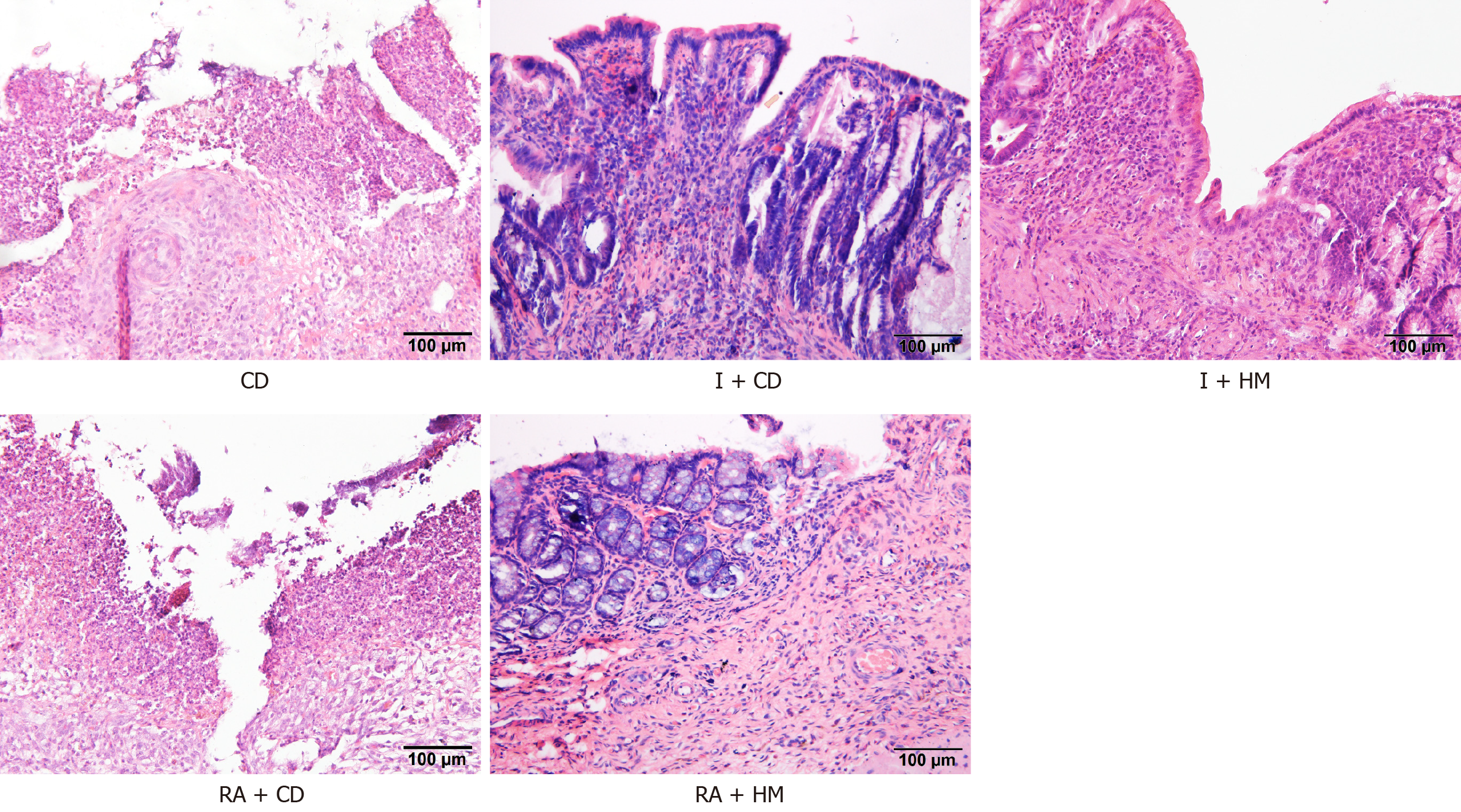
Figure 4 Histopathological observations of colon tissue from each group by hematoxylin and eosin staining.
Scale bar: 100 μm. CD: Crohn’s disease; I + CD: Insulin + Crohn’s disease; I + HM: Insulin + herbal cake-partitioned moxibustion; RA + CD: Rapamycin + Crohn’s disease; RA + HM: Rapamycin + herbal cake-partitioned moxibustion.
Figure 5 Immunofluorescence images for microtubule-associated protein 1 light chain 3 beta (Red) in each group.
Scale bar: 200 μm. CD: Crohn’s disease; I + CD: Insulin + Crohn’s disease; I + HM: Insulin + herbal cake-partitioned moxibustion; RA+CD: Rapamycin + Crohn’s disease; RA + HM: Rapamycin + herbal cake-partitioned moxibustion; LC3B: Microtubule-associated protein 1 light chain 3 beta.
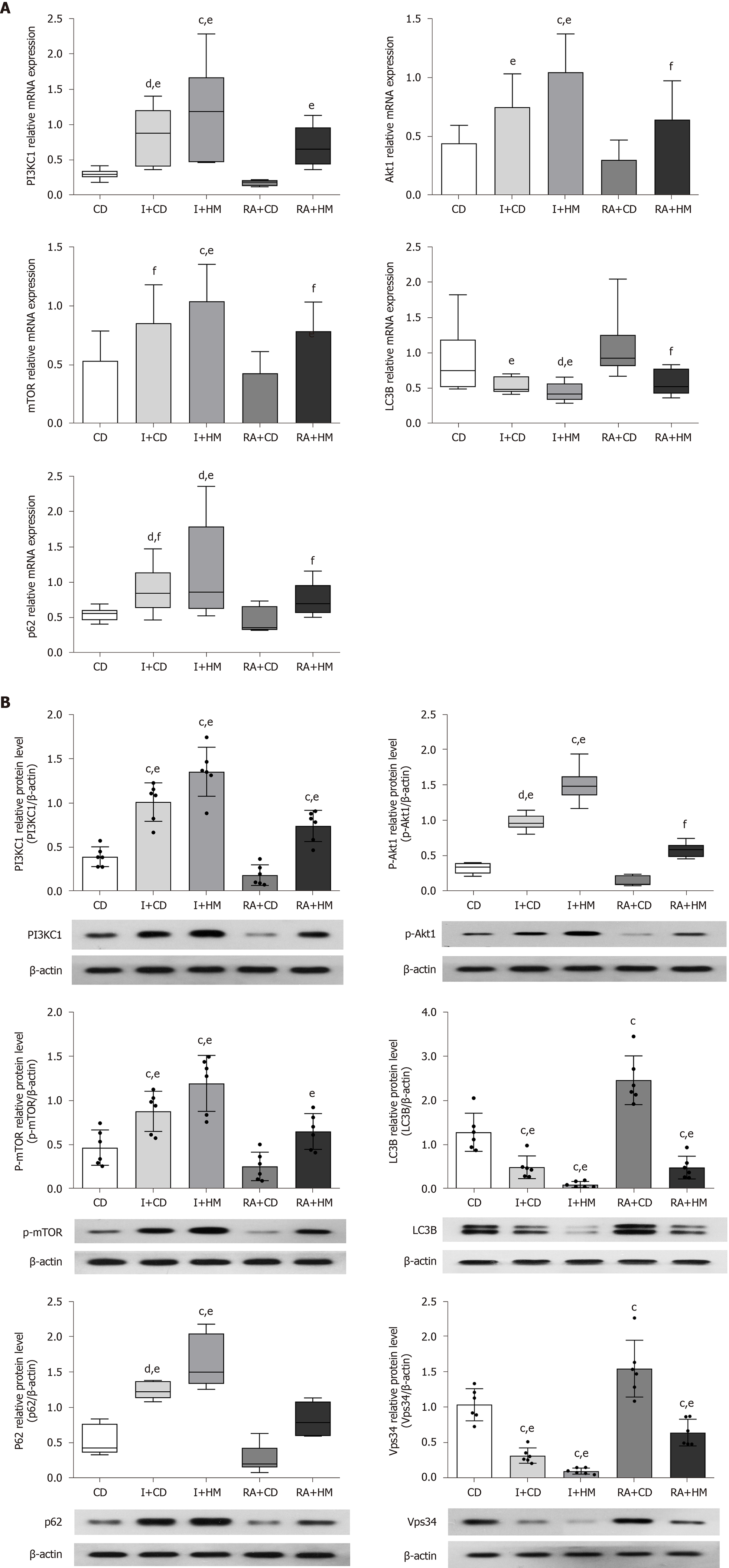
Figure 6 Herbal cake-partitioned moxibustion at the Qihai (CV6) and bilateral Tianshu (ST25) acupoints regulates the mRNA and protein expression of PI3KC signaling-related molecules in the colon tissues of Crohn’s disease rats.
A: The mRNA levels of phosphatidylinositol 3-kinase class I (PI3KC1), Akt1, mammalian target of rapamycin (mTOR), microtubule-associated protein 1 light chain 3 beta (LC3B), and sequestosome 1 (p62) were determined by real-time PCR; B: Expression changes of the PI3KC1, p-Akt1, mTOR, LC3B, p62, and class III phosphatidylinositol 3-kinase proteins evaluated by Western blot. Six independent experiments were analyzed, and the data are presented as the mean ± SD or medians (P25, P75). cP < 0.01, dP < 0.05 vs CD group; eP < 0.01, fP < 0.05 vs RA+CD group. CD: Crohn’s disease; I + CD: Insulin + Crohn’s disease; I + HM: Insulin + herbal cake-partitioned moxibustion; RA+CD: Rapamycin + Crohn’s disease; RA + HM: Rapamycin + herbal cake-partitioned moxibustion; PI3KC1: Phosphatidylinositol 3-kinase class I; Akt1: Protein kinase B akt-1; p-Akt1: Phospho-protein kinase B akt-1; mTOR: Mammalian target of rapamycin; p-mTOR: Phospho-mammalian target of rapamycin; LC3B: Microtubule-associated protein 1 light chain 3 beta; p62: Sequestosome 1; Vps34: Class III phosphatidylinositol 3-kinase.
- Citation: Wang SY, Zhao JM, Zhou CL, Zheng HD, Huang Y, Zhao M, Zhang ZY, Wu LY, Wu HG, Liu HR. Herbal cake-partitioned moxibustion inhibits colonic autophagy in Crohn’s disease via signaling involving distinct classes of phosphatidylinositol 3-kinases. World J Gastroenterol 2020; 26(39): 5997-6014
- URL: https://www.wjgnet.com/1007-9327/full/v26/i39/5997.htm
- DOI: https://dx.doi.org/10.3748/wjg.v26.i39.5997


