Copyright
©The Author(s) 2020.
World J Gastroenterol. Sep 28, 2020; 26(36): 5520-5526
Published online Sep 28, 2020. doi: 10.3748/wjg.v26.i36.5520
Published online Sep 28, 2020. doi: 10.3748/wjg.v26.i36.5520
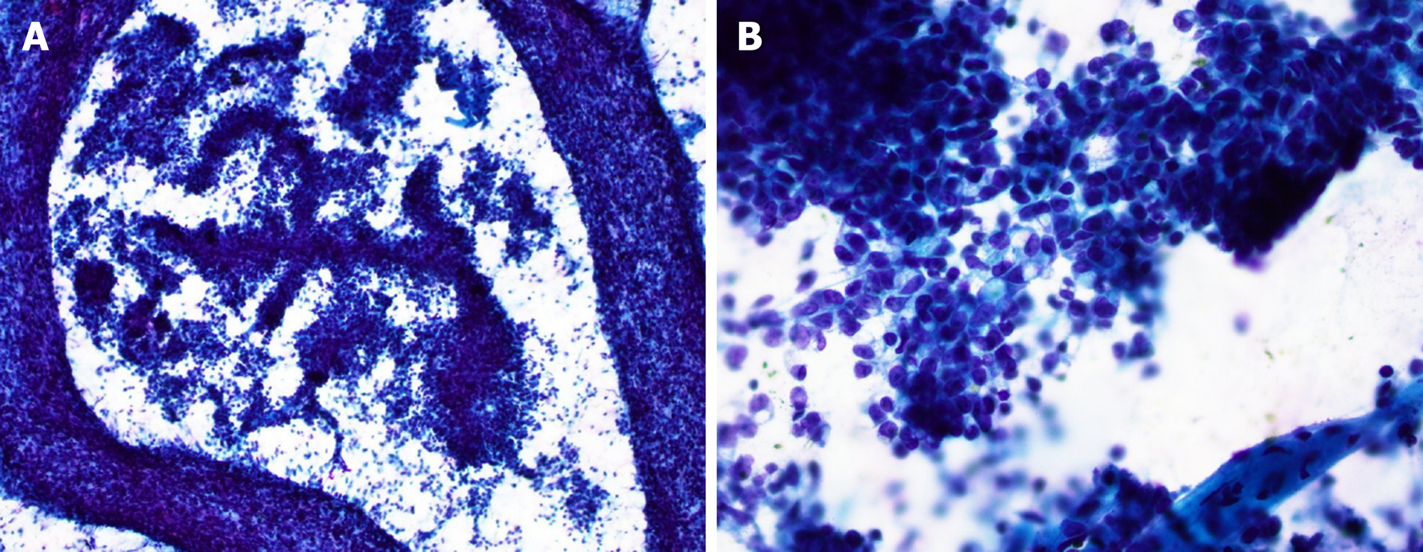
Figure 1 Fine needle aspiration smears of the pancreatic tumor.
A: Pseudopapillary growth pattern and discohesive tumor cells (Papanicolaou stain, 40 ×); B: Tumor cells with mild to moderate cytologic atypia and large intracytoplasmic vacuoles (Papanicolaou stain, 400 ×).
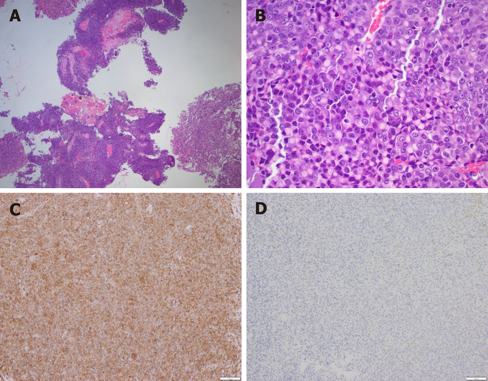
Figure 2 Histomorphology of enucleated pancreatic tumor.
A: Pseudopapillary growth pattern (Hematoxylin and eosin stain, 40 ×); B: Tumor cells with rhabdoid features (Hematoxylin and eosin stain, 400 ×); C, D: Immunohistochemistry stains of vimentin (C) and SMARCB1/INI1 (D) (100 ×).
- Citation: Hua Y, Soni P, Larsen D, Zreik R, Leng B, Rampisela D. SMARCB1/INI1-deficient pancreatic undifferentiated rhabdoid carcinoma mimicking solid pseudopapillary neoplasm: A case report and review of the literature. World J Gastroenterol 2020; 26(36): 5520-5526
- URL: https://www.wjgnet.com/1007-9327/full/v26/i36/5520.htm
- DOI: https://dx.doi.org/10.3748/wjg.v26.i36.5520


